. 2014 May 22;1(1):e11.
doi: 10.1212/NXI.0000000000000011. eCollection 2014 Jun.

AQP4 autoantibody assay performance in clinical laboratory service

Affiliations

AQP4 autoantibody assay performance in clinical laboratory service

J P Fryer et al. Neurol Neuroimmunol Neuroinflamm. .

Abstract

Objective: To compare performance of contemporary aquaporin-4-immunoglobulin (Ig) G assays in clinical service.

Methods: Sera from neurologic patients (4 groups) and controls were tested initially by service ELISA (recombinant human aquaporin-4, M1 isoform) and then by cell-based fluorescence assays: fixed (CBA, M1-aquaporin-4, observer-scored) and live (fluorescence-activated cell sorting [FACS], M1 and M23 aquaporin-4 isoforms). Group 1: all Mayo Clinic patients tested from January to May 2012; group 2: consecutive aquaporin-4-IgG-positive patients from September 2011 (Mayo and non-Mayo); group 3: suspected ELISA false-negatives from 2011 to 2013 (physician-reported, high likelihood of neuromyelitis optica spectrum disorders [NMOSDs] clinically); group 4: suspected ELISA false-positives (physician-reported, not NMOSD clinically).

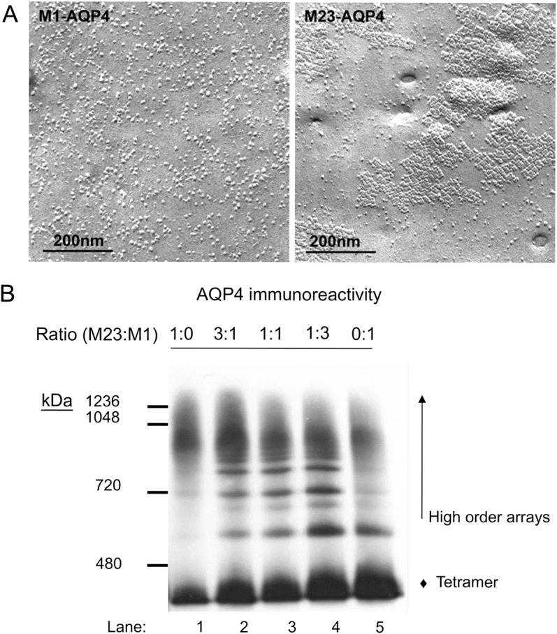
Results: Group 1 (n = 388): M1-FACS assay performed optimally (areas under the curves: M1 = 0.64; M23 = 0.57 [p = 0.02]). Group 2 (n = 30): NMOSD clinical diagnosis was confirmed by: M23-FACS, 24; M1-FACS, 23; M1-CBA, 20; and M1-ELISA, 18. Six results were suspected false-positive: M23-FACS, 2; M1-ELISA, 2; and M23-FACS, M1-FACS, and M1-CBA, 2. Group 3 (n = 31, suspected M1-ELISA false-negatives): results were positive for 5 sera: M1-FACS, 5; M23-FACS, 3; and M1-CBA, 2. Group 4 (n = 41, suspected M1-ELISA false-positives): all negative except 1 (positive only by M1-CBA). M1/M23-cotransfected cells expressing smaller membrane arrays of aquaporin-4 yielded fewer false- positive FACS results than M23-transfected cells.

Conclusion: Aquaporin-4-transfected CBAs, particularly M1-FACS, perform optimally in aiding NMOSD serologic diagnosis. High-order arrays of M23-aquaporin-4 may yield false-positive results by binding IgG nonspecifically.


Figures

Figure 1
Figure 1. Examples illustrating gating strategy for fluorescence-activated cell sorting (FACS)
Examples include (from left to right) serum from a patient with neuromyelitis optica spectrum disorder (NMOSD) yielding positivity by both M1-FACS and M23-FACS, serum from a patient with multiple sclerosis (MS) yielding positivity by M23-FACS only, and negative serum from a patient with MS. GFP = green fluorescent protein; pos = positive; pop = population.
Figure 2
Figure 2. M1 and M23 AQP4 isoforms compared by freeze-fracture electron microscopic and Western blot analyses
(A) Plasma membranes of HEK293 cells expressing recombinant M1-AQP4 or M23-AQP4 viewed by freeze-fracture electron microscopy. (B) Western blot analysis of the proportion of recombinant AQP4 expressed in higher-order arrays or as tetramers in HEK293 cells transfected with plasmids encoding M23 alone (lane 1), M1 alone (lane 5), or different ratios of each (lanes 2–4). Intramembranous particles in M1-AQP4 cells are predominantly singlet (A, left). Compare the large lattices of orthogonal array-like assemblies in M23-AQP4 cells (A, right). M1-AQP4 coexpression inhibits high-order array formation by M23-AQP4 (B). The y-axis indicates molecular weight (kDa) of AQP4 structures. Solubilized proteins, separated by Blue Native gel electrophoresis and transferred to PDF membrane, were probed with monoclonal AQP4-specific IgG. AQP4 immunoreactivity in largest-sized arrays (lane 1) diminishes with increasing M1:M23 ratio, and the proportion in tetrameric form increases (lane 5). AQP4 = aquaporin-4; Ig = immunoglobulin; HEK = human embryonic kidney.
Figure 3
Figure 3. Fluorescence-activated cell sorting (FACS) employing cells singly transfected with M1-AQP4 or M23-AQP4 or cotransfected with both AQP4 isoforms
Flow cytometry reveals nonspecific binding of control patients' serum IgG to live HEK293 cells expressing M23-AQP4, which is reduced when M1-AQP4 is coexpressed. IgG in sera of 2 positive control patients with neuromyelitis optica (NMO) binds to all AQP4-transfected cells but binds more avidly to cells expressing M23-AQP4 or both M23 and M1 (1:1 ratio) than to cells expressing M1 alone. IgG binding indices for the 14 control sera lacking NMOSD were all less than 2.00 for M1 single-transfected cells; for M23 single-transfected cells the median IgG binding index was 6.8 (range 2.98–25.8) and for M1/M23 cotransfected cells the median was 3.3 (range 1.98–14.7). The horizontal gray line indicates the cutoff for M23-FACS positivity (3.00). AQP4 = aquaporin-4; Ig = immunoglobulin; HEK = human embryonic kidney; NMOSD = NMO spectrum disorder.

References

    1. Apiwattanakul M, Popescu BF, Matiello M, et al. . Intractable vomiting as the initial presentation of neuromyelitis optica. Ann Neurol 2010;68:757–761 - PubMed
    1. Iorio R, Lucchinetti CF, Lennon VA, et al. . Syndrome of inappropriate antidiuresis may herald or accompany neuromyelitis optica. Neurology 2011;77:1644–1646 - PMC - PubMed
    1. Weinshenker BG, Wingerchuk DM, Vukusic S, et al. . Neuromyelitis optica IgG predicts relapse after longitudinally extensive transverse myelitis. Ann Neurol 2006;59:566–569 - PubMed
    1. Wingerchuk DM, Pittock SJ, Lucchinetti CF, Lennon VA, Weinshenker BG. A secondary progressive clinical course is uncommon in neuromyelitis optica. Neurology 2007;68:603–605 - PubMed
    1. Waters PJ, McKeon A, Leite MI, et al. . Serologic diagnosis of NMO: a multicenter comparison of aquaporin-4-IgG assays. Neurology 2012;78:665–671; discussion 669 - PMC - PubMed

LinkOut - more resources