. 2014 May 22;1(1):e12.
doi: 10.1212/NXI.0000000000000012. eCollection 2014 Jun.

Antibodies to MOG have a demyelination phenotype and affect oligodendrocyte cytoskeleton

Affiliations

Antibodies to MOG have a demyelination phenotype and affect oligodendrocyte cytoskeleton

Russell C Dale et al. Neurol Neuroimmunol Neuroinflamm. .

Abstract

Objective: To examine the clinical features of pediatric CNS demyelination associated with positive myelin oligodendrocyte glycoprotein (MOG) antibodies and to examine the functional effects of MOG antibody on oligodendrocyte cytoskeleton.

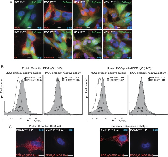
Methods: We measured MOG antibody using a fluorescence-activated cell sorting live cell-based assay in acute sera of 73 children with CNS demyelination (DEM) (median age 8 years, range 1.3-15.3) followed for a median of 4 years. We used MO3.13 cells to examine immunoglobulin (Ig) G effects on oligodendrocyte cytoskeleton using 3D deconvolution imaging.

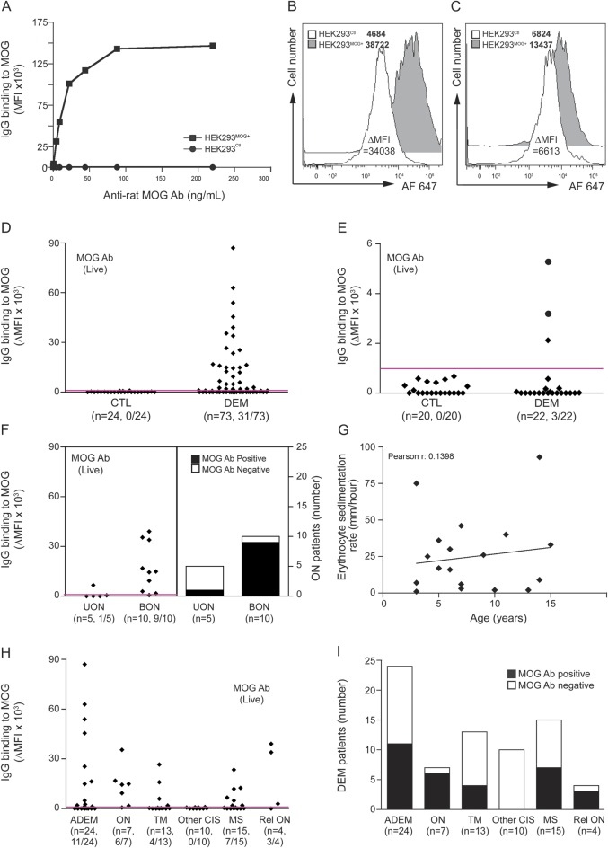
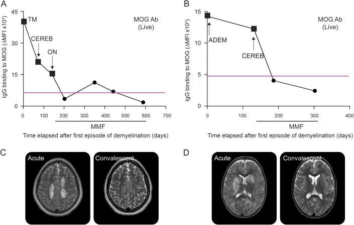
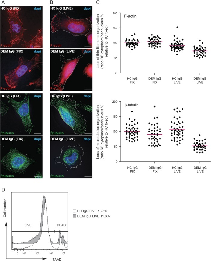
Results: MOG antibodies were found in 31/73 patients with DEM (42%) but in 0/24 controls. At first presentation, MOG antibody-positive patients were more likely to have bilateral than unilateral optic neuritis (ON) (9/10 vs 1/5, respectively, p = 0.03), less likely to have brainstem findings (2/31 vs 16/42, p = 0.005), more likely to have a raised erythrocyte sedimentation rate >20 mm/h (9/19 vs 3/21, p = 0.05), less likely to have intrathecal oligoclonal bands (0/16 vs 5/27, p = 0.18), and less likely to be homozygous or heterozygous for human leukocyte antigen DRB1*1501 (3/18 vs 7/22, p = 0.46). MOG antibody positivity varied according to clinical phenotype, with ON and relapsing ON most likely to be seropositive. Two relapsing MOG antibody-positive patients treated with mycophenolate mofetil remain in remission and have become MOG antibody seronegative. Oligodendrocytes incubated with purified IgG from MOG antibody-positive patients showed a striking loss of organization of the thin filaments and the microtubule cytoskeleton, as evidenced by F-actin and β-tubulin immunolabelings.

Conclusions: MOG antibody may define a separate demyelination syndrome, which has therapeutic implications. MOG antibody has functional effects on oligodendrocyte cytoskeleton.


Figures

Figure 1
Figure 1. Distribution of MOG IgG antibody in pediatric demyelinating diseases
(A) Antibody reactivity to myelin oligodendrocyte glycoprotein (MOG) was determined by flow cytometry live cell-based assay. (B) Representative example of flow cytometry histograms for one MOG antibody–positive patient with a very high ΔMFI and (C) an intermediate ΔMFI. MFI values are noted in the legend. (D) Human surface MOG IgG antibody was detected in 31/73 sera from patients with demyelinating diseases (DEM) and 0/24 controls (CTL). Magenta line on graph represents the positivity threshold. MOG antibody positivity is shown between brackets. (E) Surface MOG antibody was detected in 3/22 CSF from patients with DEM and 0/20 CSF from patients with other neurologic diseases (CTL). Black circles represent patients with positive MOG antibody in CSF and in serum. (F) Distribution and number of MOG antibody–positive patients in optic neuritis (ON). (G) Correlation between erythrocyte sedimentation rate and age in MOG antibody–positive patients. (H) Distribution and (I) number of MOG antibody–positive patients in demyelinating diseases at follow-up. Ab = antibody; ADEM = acute disseminated encephalomyelitis; CIS = clinically isolated syndrome; BON = bilateral optic neuritis; HEK = human embryonic kidney; Ig = immunoglobulin; MFI = mean fluorescence intensity; MS = multiple sclerosis; TM = transverse myelitis; UON = unilateral optic neuritis.
Figure 2
Figure 2. Temporal distribution of MOG antibody in serum of 2 relapsing patients with demyelinating diseases
In both patient A (A) and patient B (B), upon treatment mycophenolate mofetil (MMF), myelin oligodendrocyte glycoprotein (MOG) antibody decreased to within the healthy control range (below threshold of positivity), and these low titers were associated with remission. Representative dot plot out of 3 experiments is shown. Magenta lines on graphs represent the positivity threshold (obtained with 24 control samples). Black squares represent serum analysis during acute demyelination episodes, and black circles represent sera during remission. Type of demyelinating episode is shown on graph. (C, D) Representative T2 axial MRI scans demonstrate demyelinating lesions during the first acute event and during convalescence. Patient A (C) had globular deep white matter lesions on acute scan (left panel), which show residual gliosis on convalescent scan and no new lesions (right panel). Patient B (D) had inflammatory lesions in basal ganglia and white matter on acute scan (left panel), with complete resolution on convalescent scan and no new lesions (right panel). Ab = antibody; ADEM = acute disseminated encephalomyelitis; CEREB = cerebellar episode; Ig = immunoglobulin; MFI = mean fluorescence intensity; ON = optic neuritis; TM = transverse myelitis.
Figure 3
Figure 3. Human oligodendroglial MO3.13MOG+ cells express markers of oligodendrocytes and are immunolabeled with protein G- and human MOG-purified human IgG from MOG antibody–positive DEM patients
(A) Immunocytochemistry on fixed permeabilized cells showed that MO3.13MOG+ cells expressed oligodendrocyte markers. (B) Protein G- and human MOG-immunopurified IgG from MOG antibody–positive serum immunolabeled live HEK293MOG+ cells but did not immunolabel HEK293Ctl cells compared to MOG antibody–negative serum. Binding to cells was determined by flow cytometry. Mean fluorescence intensity (MFI) values are shown in legends. (C) Protein G- and human MOG-purified IgG from MOG antibody–positive serum also immunolabeled fixed unpermeabilized MO3.13MOG+ cells compared to MOG antibody–negative serum. Representative data are shown (volume projection of entire Z-stack acquired using 3D deconvolution microscopy). Nuclei stained with 4',6-diamidino-2-phenylindole (DAPI). Bar: 10 μm. A2B5 = c-series ganglioside-specific antigen A2B5; Ab = antibody; CNPase = 2′, 3′-cyclic nucleotide 3′-phosphodiesterase; DEM = demyelinating diseases; GalC = galactocerebroside; HEK = human embryonic kidney; Ig = immunoglobulin; MBP = myelin basic protein; MOG = myelin oligodendrocyte glycoprotein; O4 = oligodendrocyte marker O4.
Figure 4
Figure 4. Purified IgG from DEM patients induces loss of cytoskeleton organization in live human oligodendroglial MO3.13MOG+ cells
F-actin (upper images) and β-tubulin (lower images) immunolabelings in human fixed (A) or live (B) oligodendroglial MO3.13MOG+ cells incubated with purified healthy control (HC) IgG or MOG antibody–positive DEM IgG. Representative data are shown (volume projection of entire Z-stack acquired using deconvolution microscopy). Nuclei stained with 4',6-diamidino-2-phenylindole (DAPI). Bar: 10 μm. Dotted lines on right images represent contour of cells as determined by differential interference contrast images (not shown). (C) Quantification of change in distribution of thin filament (F-actin, upper scatter plot) and microtubule network (β-tubulin, lower scatter plot) organization using 3D deconvolution microscopy. F-actin and β-tubulin were majorly affected in live cells incubated with DEM IgG. Forty different cells (1 cell = 1 diamond) from 2 HC and 2 DEM patients out of 3 independent experiments are shown. Results are expressed as ratio of the relative enrichment (RE) between cytoplasm and nucleus and values shown are percentages relative to HC IgG FIX (100%). Red bars represent mean. Note that there also was a loss of organization of the thin filaments after HC IgG incubation on live cells compared to fixed conditions, but to a lesser extent than after DEM IgG. (D) Cell viability assay by flow cytometry on live human oligodendroglial MO3.13MOG+ cells incubated 45 minutes with 6 μg of purified HC IgG or DEM IgG. No difference in cell death was observed. Dead cell percentages are noted in the legend. 7AAD = 7-aminoactinomycin D; DEM = demyelinating diseases; Ig = immunoglobulin; MOG = myelin oligodendrocyte glycoprotein.

References

    1. Papadopoulos MC, Verkman AS. Aquaporin 4 and neuromyelitis optica. Lancet Neurol 2012;11:535–544 - PMC - PubMed
    1. Vincent A, Bien CG, Irani SR, Waters P. Autoantibodies associated with diseases of the CNS: new developments and future challenges. Lancet Neurol 2011;10:759–772 - PubMed
    1. Wingerchuk DM, Lennon VA, Lucchinetti CF, Pittock SJ, Weinshenker BG. The spectrum of neuromyelitis optica. Lancet Neurol 2007;6:805–815 - PubMed
    1. Johns TG, Bernard CC. The structure and function of myelin oligodendrocyte glycoprotein. J Neurochem 1999;72:1–9 - PubMed
    1. Linington C, Bradl M, Lassmann H, Brunner C, Vass K. Augmentation of demyelination in rat acute allergic encephalomyelitis by circulating mouse monoclonal antibodies directed against a myelin/oligodendrocyte glycoprotein. Am J Pathol 1988;130:443–454 - PMC - PubMed