. 2014 Oct 23;10(10):e1004666.
doi: 10.1371/journal.pgen.1004666. eCollection 2014 Oct.

Licensing of yeast centrosome duplication requires phosphoregulation of sfi1

Affiliations

Licensing of yeast centrosome duplication requires phosphoregulation of sfi1

Jennifer S Avena et al. PLoS Genet. .

Abstract

Duplication of centrosomes once per cell cycle is essential for bipolar spindle formation and genome maintenance and is controlled in part by cyclin-dependent kinases (Cdks). Our study identifies Sfi1, a conserved component of centrosomes, as the first Cdk substrate required to restrict centrosome duplication to once per cell cycle. We found that reducing Cdk1 phosphorylation by changing Sfi1 phosphorylation sites to nonphosphorylatable residues leads to defects in separation of duplicated spindle pole bodies (SPBs, yeast centrosomes) and to inappropriate SPB reduplication during mitosis. These cells also display defects in bipolar spindle assembly, chromosome segregation, and growth. Our findings lead to a model whereby phosphoregulation of Sfi1 by Cdk1 has the dual function of promoting SPB separation for spindle formation and preventing premature SPB duplication. In addition, we provide evidence that the protein phosphatase Cdc14 has the converse role of activating licensing, likely via dephosphorylation of Sfi1.


Conflict of interest statement

The authors have declared that no competing interests exist.

Figures
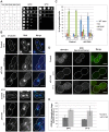
Figure 1
Figure 1. sfi1-C4A displays impaired growth and spindle and chromosome segregation defects.
A. Dilution series of strains with mutations in SFI1 on YPD. B–C. Immunofluorescent staining (α-tubulin: green, DNA: blue) of fixed large-budded sfi1-C4A (JA249) or control (JA196) cells grown at 24°C or shifted to 37°C for 8.25 h in YPD. MT: microtubule, SS: short metaphase bipolar spindle, BPS/SP: bipolar spindle or separated poles. Bar: 5 µm. C. Quantification of B. Asterisks: statistically significant difference via Student's t test between sfi1-C4A and control for each phenotypic category at 24°C (red) and 37°C (black). **p<0.01. *: p<0.05. Significance only shown for comparisons between strains at each temperature. Error bars: SD. n≥192 cells per group from 2 experiments. D–E. GFP-sfi1-C4A pLEU-HIS-SPC42-mCherry (JA354) or GFP-SFI1 pLEU-HIS-SPC42-mCherry (JA311) grown in SC-Leu at 24°C or shifted to 37°C 9 h were fixed prior to localization (D) of Sfi1 (green) and Spc42 (red) and quantification of Sfi1 levels in arbitrary units (a.u.) (E). Error bars: SD. n≥62 cells per group from 2 experiments. Bar: 5 µm.
Figure 2
Figure 2. sfi1-C4A displays unseparated SPBs or bipolar spindles with enlarged or reduplicated SPBs.
A. Asynchronous sfi1-C4A SPC42-GFP (JA302) and SFI1 SPC42-GFP (JA254) control cells grown at 24°C and shifted to 37°C for 9 h in YPD. Fixed large-budded cells were imaged by SIM. GFP on left (inset bar: 1 µm) and merge with transmitted image on right. Bar: 5 µm. B. Quantification of A. Asterisks: statistically significant difference via Student's t test between sfi1-C4A and control for each phenotypic category at 24°C (red) and 37°C (black). **p<0.01. *: p<0.05. Significance only shown for comparisons between strains at each temperature. Error bars: SD. n≥77 cells per group from 2 experiments. C. Asynchronous control cells (JA196) shifted to 37°C for 4 h in YPD and prepared for EM. Bipolar spindle with two SPBs (1, 2). Bar: 500 nm. D–F. Asynchronous sfi1-C4A (JA249) cells shifted to 37°C for 9 h in YPD and prepared for EM. Serial sections were examined by EM for 18 cells. D. Representative cell (n = 11) with two SPBs at one position. SPBs (1, 2) are at abnormal orientations to one another with a bridge. Bar: 100 nm. E–F. Multiple sections from the same cell are shown. Left panel(s): entire nucleus, 500 nm bar. Right panels: SPB(s), 100 nm bar. E. Representative cell (n = 2) containing a short bipolar spindle with SPB 1 at an orientation different from that of abnormally large SPB 2. F. Representative cell (n = 5) containing a short bipolar spindle with extra SPBs. SPB 1 is located at a region of the nuclear envelope opposite SPBs 2 and 3, which are connected via a bridge. Left panel shows the location of SPB 1, shown in the upper right panel at higher magnification, and SPBs 2 and 3. In the lower right panel, both SPBs 2 and 3 are clearly seen in a higher magnification view from a different serial section of the same cell. Average distance between adjacent SPBs: 108±10 nm.
Figure 3
Figure 3. Reduplicated SPBs upon mitotic arrest are enhanced in sfi1-C4A and are present upon Cdc14 activation.
A–C. SFI1 (JA295) or sfi1-C4A (JA297) cells containing GAL1-pds1-mdb SPC42-GFP were grown to early-log phase at 24°C in YEP with 2% raffinose then arrested in G1 with α-factor (≤2 SPBs at G1 arrest: 98.1±0.4% for sfi1-C4A, 99.4±0.4% for SFI1; n≥219 per group from 2 experiments). G1-arrested cells were released into YEP with 2% galactose to arrest in mitosis and, once at mitotic arrest, were shifted to 37°C for 2.5 h. A. Fixed large-budded cells were imaged by SIM, with GFP on left (inset bar: 1 µm) and merge with transmitted image on right. Bar: 5 µm. Mitotically-arrested cells following 37°C shift. B. Quantification of A. Asterisks: statistically significant difference via Student's t test. **p<0.01. *: p<0.05. Error bars: SD. n≥185 cells per group from 2 experiments. C. A sfi1-C4A GAL1-pds1-mdb SPC42-GFP cell imaged by EM containing at least four SPBs (1–4) total. Left panels: entire nucleus, 500 nm bar. Right: SPBs, 100 nm bar. D. An asynchronous culture of MET-CDC20 cdh1Δ GALS-ESP1 SPC42-GFP (JA256) cells was grown to early-log phase in SC-Met with 3% raffinose. Methionine was added (2 mM final concentration) to arrest cells in metaphase and every 2 h for the experiment remainder. After metaphase arrest, Esp1 either was induced for 4 h using 3% galactose to release Cdc14 from the nucleolus or remained uninduced in raffinose. Fixed large-budded cells were imaged by SIM, with GFP on left (inset bar: 1 µm) and merge with transmitted image on right. Bar: 5 µm. n≥211 per group from 2 experiments. Upper panel: cells with no Esp1 induction with bipolar spindles (63±13%). Lower panels: cells with Esp1 induction with one or two reduplicated SPBs (3 or 4 Spc42-GFP foci total, respectively; 22±1%); p = 0.02 (Student's t test) for reduplicated SPBs versus without Esp1 induction (7±3%).
Figure 4
Figure 4. Model for licensing of yeast centrosome duplication via phosphoregulation of Sfi1.
Cdk1 activity is required for SPB separation, in which Cdk1 phosphorylates the Sfi1 C terminus, ensuring SPB duplication does not begin until completion of mitosis and downregulation of mitotic cyclin/Cdk1. Dephosphorylation of Sfi1, likely by Cdc14, licenses SPB duplication to allow the next cycle of SPB duplication to begin at G1. cMT: cytoplasmic microtubules. nMT: nuclear microtubules. NE: nuclear envelope. ▪: satellite.

Similar articles

Cited by

References

    1. Ganem NJ, Godinho SA, Pellman D (2009) A mechanism linking extra centrosomes to chromosomal instability. Nature 460: 278–282 10.1038/nature08136 - DOI - PMC - PubMed
    1. Haase SB, Winey M, Reed SI (2001) Multi-step control of spindle pole body duplication by cyclin-dependent kinase. Nat Cell Biol 3: 38–42 10.1038/35050543 - DOI - PubMed
    1. Vidwans SJ, Wong ML, O'Farrell PH (2003) Anomalous centriole configurations are detected in Drosophila wing disc cells upon Cdk1 inactivation. J Cell Sci 116: 137–143. - PMC - PubMed
    1. Tsou M-FB, Stearns T (2006) Mechanism limiting centrosome duplication to once per cell cycle. Nature 442: 947–951 10.1038/nature04985 - DOI - PubMed
    1. Tsou M-FB, Wang W-J, George KA, Uryu K, Stearns T, et al. (2009) Polo kinase and separase regulate the mitotic licensing of centriole duplication in human cells. Dev Cell 17: 344–354 10.1016/j.devcel.2009.07.015 - DOI - PMC - PubMed

Publication types

MeSH terms