. 2014 Oct 23;10(10):e1004466.
doi: 10.1371/journal.ppat.1004466. eCollection 2014 Oct.

HPV16-E7 expression in squamous epithelium creates a local immune suppressive environment via CCL2- and CCL5- mediated recruitment of mast cells

Affiliations

HPV16-E7 expression in squamous epithelium creates a local immune suppressive environment via CCL2- and CCL5- mediated recruitment of mast cells

Anne-Sophie Bergot et al. PLoS Pathog. .

Abstract

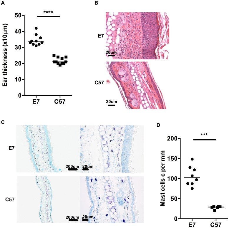
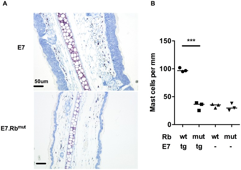
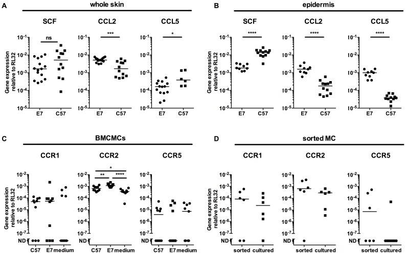
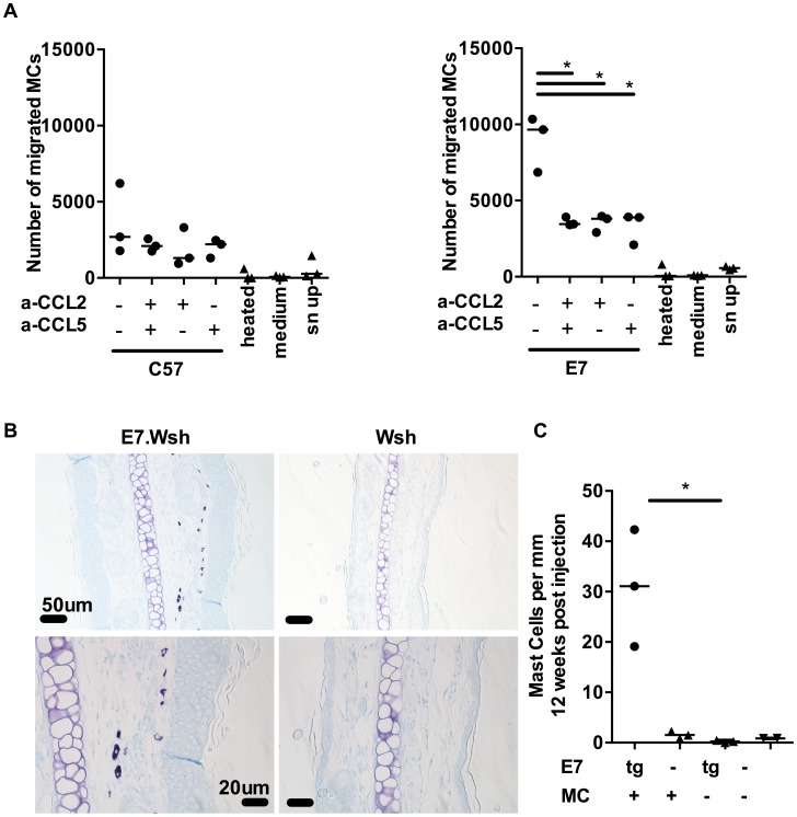
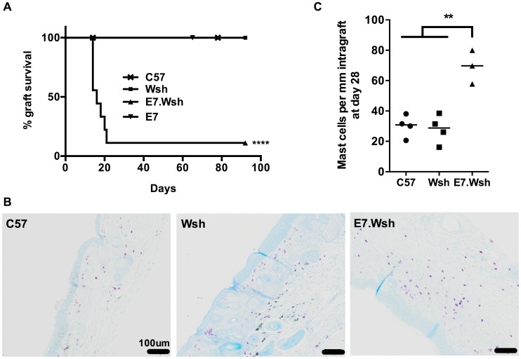
Human Papillomavirus (HPV) 16 E7 protein promotes the transformation of HPV infected epithelium to malignancy. Here, we use a murine model in which the E7 protein of HPV16 is expressed as a transgene in epithelium to show that mast cells are recruited to the basal layer of E7-expressing epithelium, and that this recruitment is dependent on the epithelial hyperproliferation induced by E7 by inactivating Rb dependent cell cycle regulation. E7 induced epithelial hyperplasia is associated with increased epidermal secretion of CCL2 and CCL5 chemokines, which attract mast cells to the skin. Mast cells in E7 transgenic skin, in contrast to those in non-transgenic skin, exhibit degranulation. Notably, we found that resident mast cells in E7 transgenic skin cause local immune suppression as evidenced by tolerance of E7 transgenic skin grafts when mast cells are present compared to the rejection of mast cell-deficient E7 grafts in otherwise competent hosts. Thus, our findings suggest that mast cells, recruited towards CCL2 and CCL5 expressed by epithelium induced to proliferate by E7, may contribute to an immunosuppressive environment that enables the persistence of HPV E7 protein induced pre-cancerous lesions.


Conflict of interest statement

The authors have declared that no competing interests exist.

Figures

Figure 1
Figure 1. MCs are highly represented in E7 skin.
A) Ear thicknesses were measured in naive age-matched E7 and C57 mice using a micrometer gauge (n = 10 and 11, respectively, ****p<0.0001). B) Representative images of E7 and C57 ear skin analyzed by H&E stain (scale bar  = 20 µm). C) Representative images of E7 and C57 ear skin analyzed by toluidine blue stain, which is specific for MCs (purple, scale bar  = 100 µm (left) and 20 µm (right)). D) MC numbers expressed per mm cartilage length (n = 8 E7 mice and n = 6 C57 mice, respectively; ***p<0.001).
Figure 2
Figure 2. MC infiltration in ear skin is dependent on E7 hyperplasia.
E7.Rbmut ear skin is not hyperplasic and contains normal MC numbers. (A) Representative images of E7.Rbwt and E7.Rbmut ear skin by toluidine blue stain (original magnification ×400, scale  = 50 µm). (B) MC number per mm cartilage length (n = 3 in each group). ***p<0.001 by unpaired t-test for indicated comparison. Rbwt are same as C57.
Figure 3
Figure 3. E7 epidermis produce CCL2 and CCL5 and MCs express the corresponding receptors.
(A) C57 and E7 whole skin, or (B) C57 and E7 epidermis, were analyzed for SCF, CCL5 (Rantes) and CCL2 (MCP-1) gene expression. Data are combined from 2 independent experiments. CCR1 and CCR5 (two Rantes receptors) and CCR2 (MCP-1 receptor) gene expression in (C) BMCMCs after 72 h culture in E7 or C57 ear skin culture supernatant or medium (n = 10 different batches of BMCMCs; data are combined from 4 independent experiments. ND, not detectable) and (D) Mast cells sorted from E7 ear skin (n = 6 independent experiments, each with 4 or 5 mice pooled per treatment). Gene expression was performed by real time-PCR relative to the RL32 housekeeping gene. *p<0.05; **p<0.01; ***p<0.001; or ****p<0.0001 by unpaired t-test for indicated comparisons; ns  =  not significant.
Figure 4
Figure 4. MCs migrate towards CCL2 and CCL5 and are recruited to E7 ear skin.
(A) 5 µm transwell assays were used to determine the mast cells chemotaxis towards chemokines. Graphs represent the number of migrated FcεRIα+ cKit+ BMCMCs towards C57 skin explant supernatants (left) or E7 explant supernatants (right) in the presence of anti-CCL2 and/or anti-CCL5 blocking antibodies used at 10 µg/ml. 3 independent experiments (B,C) Kit W-sh/W-sh (Wsh) (n = 3) and E7.Kit W-sh/W-sh (E7.Wsh) (n = 3) mice were reconstituted with 1.4×10⁷ BMCMCs i.v. or left untreated (E7.Wsh (n = 3) and Wsh (n = 2)). 12 weeks later, mice were culled and MCs were identified in ear skin by (B) toluidine blue staining (top, scale bar  = 50 µm and bottom, scale bar  = 20 µm) and expressed as (C) MC number per mm cartilage length. *p<0.01, by unpaired t-test for all indicated comparisons.
Figure 5
Figure 5. MCs are immunosuppressive in E7 environment.
(A) C57 mice were double grafted with C57 syngeneic ear skin (n = 9), and either Kit W-sh/W-sh (Wsh) ear skin (n = 9) or E7. Kit W-sh/W-sh (E7.Wsh) ear skin (n = 9). Control E7 ear skin (n = 6) are tolerated. Kaplan-Meier survival curves show the median graft survival. ****p<0.0001 using a log-rank test. Data are combined from 3 independent experiments. (B) Representative graft histology of C57, Wsh and one remaining E7.Wsh graft at day 90 by toluidine blue staining (scale bar  = 100 µm). (C) MCs per mm of graft tissue at day 28 in C57 (n = 4), Wsh non-rejected (n = 4) and E7.Wsh rejected (n = 3) grafts. (**p<0.01, by unpaired t-test).

References

    1. Woods RS, O'Regan EM, Kennedy S, Martin C, O'Leary JJ, et al. (2014) Role of human papillomavirus in oropharyngeal squamous cell carcinoma: A review. World J Clin Cases 2: 172–193. - PMC - PubMed
    1. Frazer IH, Leggatt GR, Mattarollo SR (2011) Prevention and treatment of papillomavirus-related cancers through immunization. Annu Rev Immunol 29: 111–138. - PubMed
    1. Galli SJ, Tsai M (2012) IgE and mast cells in allergic disease. Nat Med 18: 693–704. - PMC - PubMed
    1. Sayed BA, Christy A, Quirion MR, Brown MA (2008) The master switch: the role of mast cells in autoimmunity and tolerance. Annu Rev Immunol 26: 705–739. - PubMed
    1. Kobayashi A, Greenblatt RM, Anastos K, Minkoff H, Massad LS, et al. (2004) Functional attributes of mucosal immunity in cervical intraepithelial neoplasia and effects of HIV infection. Cancer Res 64: 6766–6774. - PubMed

Publication types

Substances

LinkOut - more resources