. 2014 Oct 23;10(10):e1004417.
doi: 10.1371/journal.ppat.1004417. eCollection 2014 Oct.

A novel function of human Pumilio proteins in cytoplasmic sensing of viral infection

Affiliations

A novel function of human Pumilio proteins in cytoplasmic sensing of viral infection

Ryo Narita et al. PLoS Pathog. .

Abstract

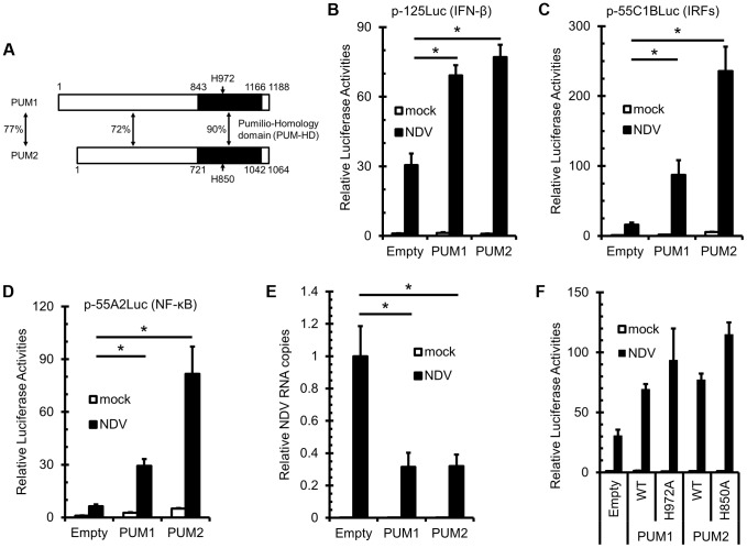
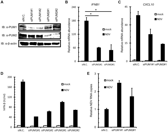
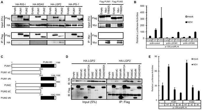
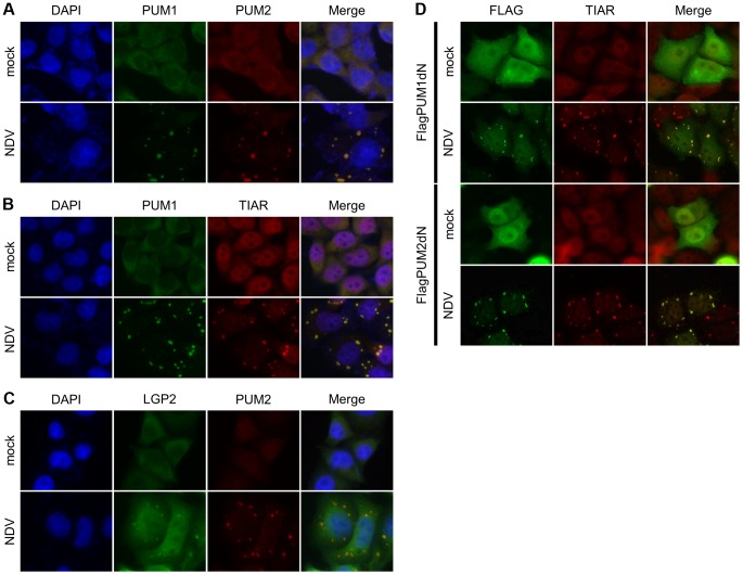
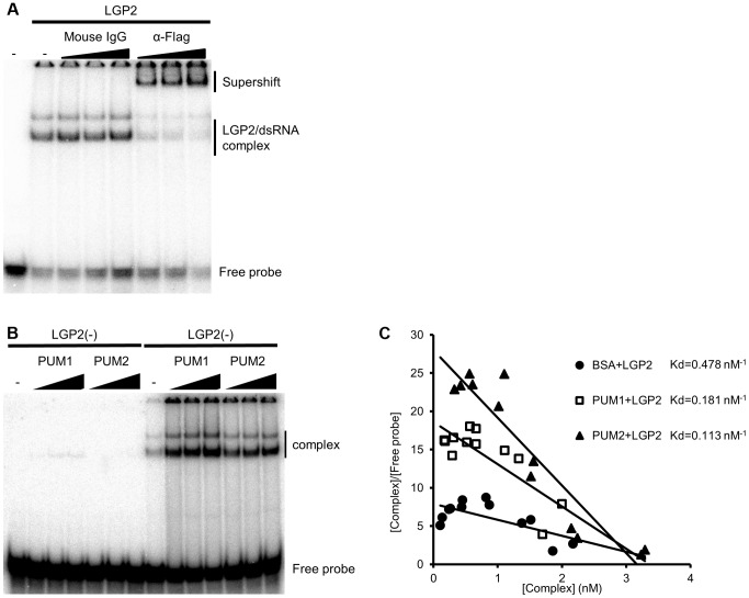
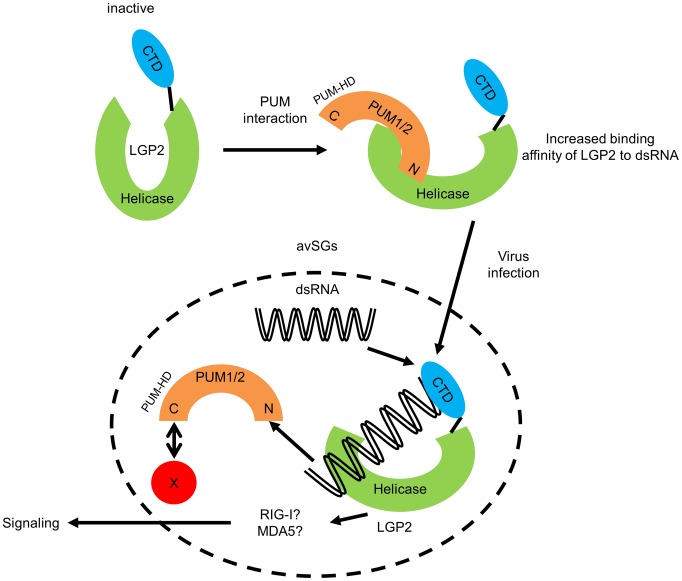
RIG-I-like receptor (RLR) plays a pivotal role in the detection of invading pathogens to initiate type I interferon (IFN) gene transcription. Since aberrant IFN production is harmful, RLR signaling is strictly regulated. However, the regulatory mechanisms are not fully understood. By expression cloning, we identified Pumilio proteins, PUM1 and PUM2, as candidate positive regulators of RIG-I signaling. Overexpression of Pumilio proteins and their knockdown augmented and diminished IFN-β promoter activity induced by Newcastle disease virus (NDV), respectively. Both proteins showed a specific association with LGP2, but not with RIG-I or MDA5. Furthermore, all of these components were recruited to NDV-induced antiviral stress granules. Interestingly, biochemical analyses revealed that Pumilio increased double-stranded (ds) RNA binding affinity of LGP2; however, Pumilio was absent in the dsRNA-LGP2 complex, suggesting that Pumilio facilitates viral RNA recognition by LGP2 through its chaperon-like function. Collectively, our results demonstrate an unknown function of Pumilio in viral recognition by LGP2.


Conflict of interest statement

The authors have declared that no competing interests exist.

Figures

Figure 1
Figure 1. Overexpression of PUM1 and PUM2 results in enhanced NDV-induced IFNB promoter activity.
(A) Schematic representation of PUM1 and PUM2. PUM-HD shows high sequence similarity between PUM1 and PUM2. Positions of histidine (H) residues critical for NRE recognition are indicated. (B–D) L929 cells were transfected with the indicated reporter gene, p-125Luc (B), p-55C1BLuc (C) or p-55A2Luc (D), and pRL-tk, together with the expression vector for PUM1 or PUM2. The cells were stimulated by NDV infection for 9 h and subjected to a dual-luciferase assay. (E) L929 cells were transfected with an expression vector for PUM1 or PUM2. The cells were infected with NDV for 24 h, and then NDV RNA levels were determined by quantitative RT-PCR. (F) L929 cells were transfected with p-125Luc and pRL-tk, together with the expression vector for wt and histidine mutants of PUM1 or PUM2 as indicated. The cells were stimulated by NDV infection and subjected to a dual-luciferase assay. Data are from one representative of at least two independent experiments; means and S.D. of duplicate experiments are shown (*p<0.05).
Figure 2
Figure 2. Knockdown of PUM1 and PUM2 downregulates NDV-induced gene activation.
(A–C) HEK293T cells were transfected with control siRNA (siN.C.) or siRNA targeting human PUM1 or PUM2 for 48 h. Knockdown efficiency was confirmed by immunoblotting with anti-PUM1, anti-PUM2 and anti-β-actin antibodies (A). The cells were infected with NDV for 9 h, and IFNB (B) and CXCL10 (C) mRNA levels were determined by quantitative RT-PCR. (D and E) HEK293T cells were transfected with control siRNA or siRNA targeting PUM1 or PUM2 for 48 h. The cells were infected with NDV for 24 h. The culture media were collected and subjected to IFN-β ELISA (D). Total cellular RNA was extracted and subjected to qRT-PCR for NDV RNA (E). Data are from one representative of at least two independent experiments; means and S.D. of duplicate experiments are shown (*p<0.05, **p<0.01).
Figure 3
Figure 3. Physical association of PUM1 and PUM2 with LGP2 and involvement of N- and C-terminal domains of PUM1 and PUM2 in IFN induction.
(A) HEK293T cells were transfected with expression vector HA-tagged RIG-I, MDA5, LGP2 or IPS-1, together with Flag-tagged PUM1 or PUM2. For TRIM25, HEK293T cells were transfected with Flag-tagged PUM1 or PUM2, together with Myc-tagged TRIM25. The cell lysates were subjected to anti-Flag or anti-c-Myc immunoprecipitation (IP), followed by Western blotting. Western blotting result of total lysate is shown as a reference (Input, 5%). (B) L929 cells were transfected with control shRNA construct (pU6i-control) or shRNA for LGP2 (pU6i-LGP2#1 and #2) and expression vectors for PUM1 or PUM2 and p-125Luc reporter and pRLtk as indicated. The cells were stimulated by infection with NDV for 9 h and subjected to the dual luciferase assay. (C) Schematic representation of PUM1 or PUM2 deletion mutants used for IP experiments. (D) HEK293T cells were transfected with expression vectors for the indicated proteins and for HA-tagged LGP2. The cell lysates were subjected to IP with anti-Flag, followed by immunoblotting with anti-HA. Immunoblotting result of total lysate is shown as a reference (Input, 5%). (E) L929 cells were transfected with expression vectors for the wild type or mutant of PUM1 or PUM2 and p-125Luc reporter and pRL-tk as indicated. Cells were stimulated by infection with NDV for 9 h and subjected to the dual luciferase assay. Data are from one representative of at least two independent experiments (means and s.d. of duplicate experiments.)
Figure 4
Figure 4. Cellular localization of PUM1, PUM2 and LGP2.
(A–C) HeLa cells were mock-treated or infected with NDV for 9 h, fixed and stained with the indicated antibodies. Nuclei were stained with DAPI. (D) HEK293T cells were transfected with the expression vector for Flag-PUM1dN or Flag-PUM2dN for 48 h and mock treated or infected with NDV for 9 h. The cells were stained with anti-Flag or anti-TIAR.
Figure 5
Figure 5. In vitro binding assay of dsRNA and LGP2.
(A and B) Recombinant LGP2 proteins (0.125 µg) were mixed with 32P-labeled dsRNA in the presence of a control mouse IgG or anti-Flag antibody (0.1, 0.2 or 0.4 µg) (A) or in the presence or absence of Pumilio proteins (0.1, 0.3 or 0.5 µg) (B). The mixture were separated by acrylamide gel and the radioactivity was analyzed. (C) LGP2 dsRNA binding affinities in the absence (filled circles) or presence of PUM1 (open square) or PUM2 (filled triangle) were analyzed and the Kd values were determined.
Figure 6
Figure 6. Hypothetical model for regulation of LGP2 by PUM1 and PUM2 in avSG.
N-terminal domain of PUM1 and PUM2 possess intrinsic affinity to LGP2. This interaction confers higher binding affinity of LGP2 to viral dsRNA. Conformational change of LGP2 is one of the explanations for the increased affinity. Viral infection such as NDV induces avSGs and accumulation of viral dsRNA, LGP2, PUM1, PUM2 and other avSG markers into avSGs. Within avSG, dsRNA interacts with LGP2/PUM complex, producing LGP2/dsRNA complex and Pumilio proteins are released from the complex. Then, LGP2 triggers signals presumably in cooperation with RIG-I or MDA5. X: potential interacting partner of C-terminal domain of PUM1 and PUM2 determining their avSG localization.

References

    1. Kawai T, Akira S (2011) Toll-like receptors and their crosstalk with other innate receptors in infection and immunity. Immunity 34: 637–650. - PubMed
    1. Yoneyama M, Fujita T (2009) RNA recognition and signal transduction by RIG-I-like receptors. Immunological reviews 227: 54–65. - PubMed
    1. Yan N, Chen ZJ (2012) Intrinsic antiviral immunity. Nature immunology 13: 214–222. - PMC - PubMed
    1. Ireton RC, Gale M Jr (2011) RIG-I like receptors in antiviral immunity and therapeutic applications. Viruses 3: 906–919. - PMC - PubMed
    1. Schlee M, Hartmann G (2010) The chase for the RIG-I ligand—recent advances. Molecular therapy: the journal of the American Society of Gene Therapy 18: 1254–1262. - PMC - PubMed

Publication types

MeSH terms