Review
. 2015:77:29-55.
doi: 10.1146/annurev-physiol-021014-071622. Epub 2014 Oct 17.

Mechanisms of ventricular arrhythmias: from molecular fluctuations to electrical turbulence

Affiliations
Review

Mechanisms of ventricular arrhythmias: from molecular fluctuations to electrical turbulence

Zhilin Qu et al. Annu Rev Physiol. 2015.

Abstract

Ventricular arrhythmias have complex causes and mechanisms. Despite extensive investigation involving many clinical, experimental, and computational studies, effective biological therapeutics are still very limited. In this article, we review our current understanding of the mechanisms of ventricular arrhythmias by summarizing the state of knowledge spanning from the molecular scale to electrical wave behavior at the tissue and organ scales and how the complex nonlinear interactions integrate into the dynamics of arrhythmias in the heart. We discuss the challenges that we face in synthesizing these dynamics to develop safe and effective novel therapeutic approaches.

Keywords: chaos; multiscale dynamics; nonlinear dynamics; sudden cardiac death; ventricular arrhythmias.


Figures

Figure 1
Figure 1
Subcellular and cellular dynamics of ventricular myocytes. (a) A single ion channel opening and closing stochastically. (b) Normal action potentials and Ca2+ transient for endocardial myocytes (left) and epicardial myocytes (right). A spike-and-dome morphology occurs in epicardial myocytes. (c) A Ca2+ release unit (CRU) that is composed of a ryanodine receptor (RyR) cluster in the sarcoplasmic reticulum (SR) membrane and an L-type Ca2+ channel (LCC) cluster in the apposing T-tubule (TT) membrane (left) and a Ca2+ spark (right). The spark image was downloaded from https://sites.google.com/site/sparkmasterhome/faq. (d ) A planar Ca2+ wave (left; arrows indicate the direction of propagation, and the numbers indicate time in milliseconds) from a Purkinje cell (from Reference with permission) and a spiral Ca2+ wave (right) (from Reference with permission). (e) Early afterdepolarizations (EADs), delayed afterdepolarizations (DADs), and triggered activity (TA). (f) Action potential alternans. ( g) Spontaneous oscillations (automaticity).
Figure 2
Figure 2
Tissue-scale excitation dynamics. (a) A rectilinear (planar) wave. (b) A focal excitation (target wave). (c) Reentry around an obstacle. (d ) A spiral wave. (e) A scroll wave. (f) A scroll wave in the ventricles (from Reference with permission). Voltage levels are indicated by the color bar, and arrows indicate the directions of propagation in panels ae. In panel f, only voltage higher than a certain value is colored in red for three-dimensional visualization.
Figure 3
Figure 3
Mechanisms of reentry initiation. (a) Reentry around an anatomic obstacle. (i ) Homogeneous refractoriness: A premature ventricular complex (PVC) successfully propagates through both sides of the obstacle without forming reentry. (ii ) Heterogeneous refractoriness: The refractory period in one region is longer than elsewhere (e.g., the gray zone in the right pathway) such that a properly timed PVC is blocked in the right pathway, propagates successfully through the contralateral pathway (e.g., the left), and then reenters the right pathway from the retrograde direction, initiating reentry around the obstacle (red arrows). (iii ) Narrow pathway: The right pathway has a very narrow exit (indicated by the circle) such that a PVC cannot conduct out of the pathway, but the impulse from outside can enter the pathway, resulting in reentry (red arrows). (b) Reentry in heterogeneous tissue in which the central region has a longer refractory period than the rest of the tissue. (c) Reentry induction by a trigger and substrate from the same source. Cells in the central region exhibit early afterdepolarizations.
Figure 4
Figure 4
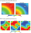
Dynamically induced dispersion of refractoriness. (a) Spatially discordant action potential duration (APD) alternans in which APD is short in one region but long in another region. In the following beat, the spatial APD pattern is reversed. (b) Spatiotemporal chaotic dynamics in the presence of early afterdepolarizations (EADs). The APD distribution in tissue exhibits an irregular spatial pattern and varies from beat to beat. EADs occur in long-APD regions (red ), but not in short-APD regions (blue).
Figure 5
Figure 5
Schematic diagram of ionic currents, transporters, signaling pathways, and their interactions in a ventricular myocyte. A dashed line with arrow indicates that signaling enhances activity, and a dashed line with bar indicates suppression of activity. See text for details.

References

    1. Janse MJ, Rosen MR. History of arrhythmias. Handb Exp Pharmacol. 2006;2006:1–39. - PubMed
    1. Jalife J. Ventricular fibrillation: mechanisms of initiation and maintenance. Annu Rev Physiol. 2000;62:25–50. - PubMed
    1. Zipes DP, Wellens HJ. Sudden cardiac death. Circulation. 1998;98:2334–51. - PubMed
    1. Priori SG, Napolitano C. Genetics of cardiac arrhythmias and sudden cardiac death. Ann N Y Acad Sci. 2004;1015:96–110. - PubMed
    1. Marsman RF, Tan HL, Bezzina CR. Genetics of sudden cardiac death caused by ventricular arrhythmias. Nat Rev Cardiol. 2014;11:96–111. - PubMed

MeSH terms