Review
. 2014 Oct 22;15(10):19203-25.
doi: 10.3390/ijms151019203.

Glutathionylation of the L-type Ca2+ channel in oxidative stress-induced pathology of the heart

Affiliations
Review

Glutathionylation of the L-type Ca2+ channel in oxidative stress-induced pathology of the heart

Victoria P A Johnstone et al. Int J Mol Sci. .

Abstract

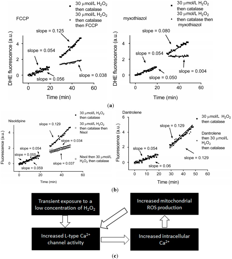
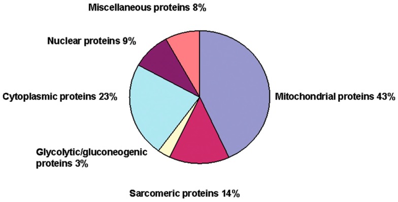
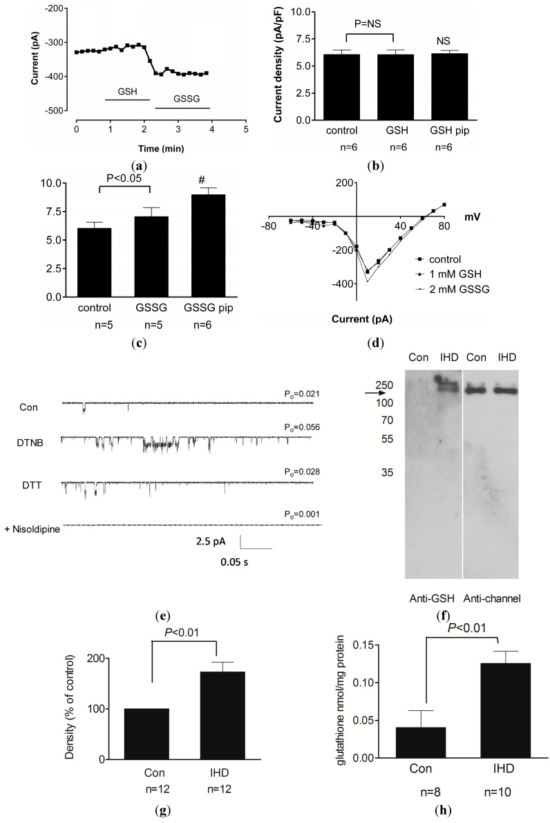
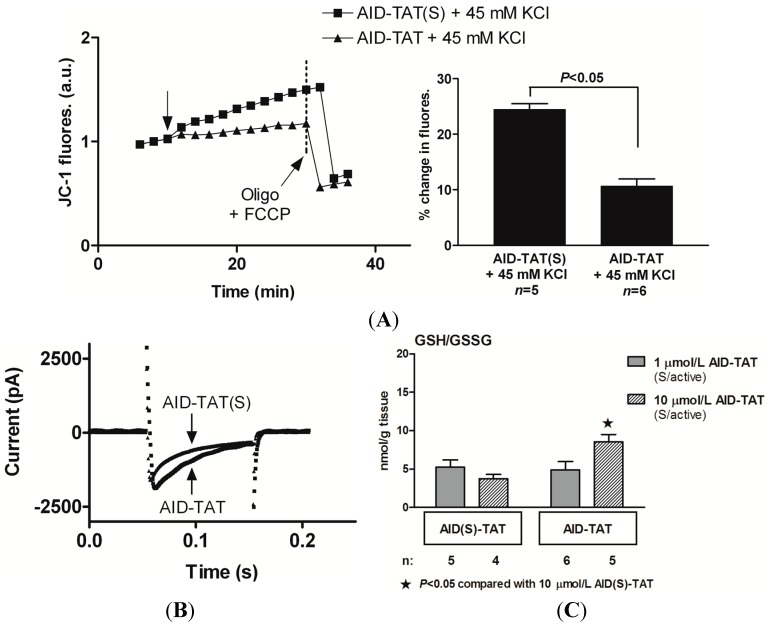
There is mounting evidence to suggest that protein glutathionylation is a key process contributing to the development of pathology. Glutathionylation occurs as a result of posttranslational modification of a protein and involves the addition of a glutathione moiety at cysteine residues. Such modification can occur on a number of proteins, and exerts a variety of functional consequences. The L-type Ca2+ channel has been identified as a glutathionylation target that participates in the development of cardiac pathology. Ca2+ influx via the L-type Ca2+ channel increases production of mitochondrial reactive oxygen species (ROS) in cardiomyocytes during periods of oxidative stress. This induces a persistent increase in channel open probability, and the resulting constitutive increase in Ca2+ influx amplifies the cross-talk between the mitochondria and the channel. Novel strategies utilising targeted peptide delivery to uncouple mitochondrial ROS and Ca2+ flux via the L-type Ca2+ channel following ischemia-reperfusion have delivered promising results, and have proven capable of restoring appropriate mitochondrial function in myocytes and in vivo.


Figures

Figure 1
Figure 1
Transient exposure to H2O2 leads to L-type Ca2+ channel-dependent increase in mitochondrial superoxide, triggering ROS-induced-ROS release. (a) dihydroethidium (DHE) fluorescence recorded from individual guinea pig cardiomyocytes before and after exposure to 30 µM H2O2 for 5 min followed by 10 U/mL catalase for 5 min then 2 nM FCCP (an uncoupler of oxidative phosphorylation) as indicated (left panel) or 7 nM myxothiazol (to block electron transport at mitochondrial complex III) (right panel); (b) DHE fluorescence recorded from individual guinea pig cardiomyocytes before and after exposure to 30 µM H2O2 for 5 min followed by 10 U/mL catalase for 5 min and the L-type Ca2+ channel antagonist nisoldipine (nisol; 2 µM) as indicated (left panel) or ryanodine receptor antagonist dantrolene (20 µM; right panel); and (c) Schematic illustrating the persistent increase in intracellular ROS and intracellular Ca2+ following a transient H2O2 exposure or ROS-induced ROS-release. Reproduced with permission from [40].
Figure 2
Figure 2
Transient exposure of myocytes to H2O2 alters mitochondrial protein synthesis. Exposure of guinea-pig myocytes to 30 µM H2O2 for 5 min followed by 10 U/mL catalase for 5 min is sufficient to induce alterations in cellular protein synthesis consistent with the development of myocyte hypertrophy. iTRAQ-facilitated proteomics analysis indicated that the majority of changes in protein synthesis are mitochondrial in origin. For further details see text. Reproduced with permission from [50].
Figure 3
Figure 3
Oxidative stress causes persistent enhancement of the L-type Ca2+ channel via ROS-mediated glutathionylation. (a) Time course of changes in membrane current recorded from guinea pig cardiac myocytes during extracellular exposure to 1 mM GSH followed by 2 mM GSSG as indicated; (b) Mean (±SEM) of L-type Ca2+ channel current density under control conditions (no drugs) and after exposure to GSH applied either extracellularly or in the patch pipette (GSH pip). NS, not significant; (c) Mean (±SEM) of L-type Ca2+ channel current density under control conditions (no drugs) and after exposure to GSSG applied either extracellularly or in the patch pipette (GSSG pip). #, p < 0.05 vs. control; (d) Current-voltage relationship for representative myocytes during voltage steps from −60 to +80 mV and exposure to GSH or GSSG as indicated; (e) Representative single channel currents recorded at −100 mV in the absence and presence of 200 µM DTNB followed by 1 mM DTT and then 2 µM nisoldipine. The open probability (P0) for each treatment is indicated; (f) Immunoblot demonstrating glutathionylation of protein after probing with anti-GSH antibody (left) and anti-channel antibody (right) in immunoprecipitated Cav1.2 channel protein samples from control nonischemic human heart (Con) and ischemic human heart (IHD); (g) Densitometry analysis of immunoblots for glutathionylated protein band normalised to the channel protein in the same lane for Con samples and IHD samples (mean ± SEM); and (h) Concentration of protein-glutathione mixed-disulfides in channel protein from Con and IHD heart samples (mean ± SEM). Reproduced with permission from [74].
Figure 4
Figure 4
Targeted peptide delivery disrupts cross-talk between the L-type Ca2+ channel and the mitochondria and reduces ischemia-reperfusion injury. (A) Exposure to AID–TAT attenuates the increase in mitochondrial membrane potential (Ψm) associated with activation of the channel. Ψm assessed as changes in JC-1 fluorescence recorded from two representative guinea pig myocytes before and after exposure to either 1 µM scrambled AID–TAT (AID–TAT(S)) or 1 µM AID–TAT followed by 45 mM KCl to activate the L-type Ca2+ channel. Arrow indicates where KCl was added. Mean ± SEM of changes in JC-1 fluorescence for myocytes exposed to AID–TAT(S) and AID–TAT shown on the right; (B) L-type Ca2+ channel current traces activated by voltage step to +10 mV from a holding potential of −30 mV from representative myocytes that were pre-incubated with either AID–TAT(S) or AID–TAT; and (C) GSH/GSSG ratio of guinea pig hearts treated with 1 or 10 µM scrambled AID–TAT (AID(S)–TAT) or active AID–TAT within 5 min after commencement of reperfusion following 30 min of no-flow ischemia. Panels A and B reproduced with permission from [104] and panel C reproduced with permission from [114].

References

    1. Dhalla N.S., Temsah R.F., Netticadan T. Role of oxidative stress in cardiovascular diseases. J. Hyperten. 2000;18:655–673. - PubMed
    1. Turrens J.F. Mitochondrial formation of reactive oxygen species. J. Physiol. 2003;552:335–344. - PMC - PubMed
    1. Schafer F.Q., Buettner G.R. Redox environment of the cell as viewed through the redox state of the glutathione disulfide/glutathione couple. Free Radic. Biol. Med. 2001;30:1191–1212. doi: 10.1016/S0891-5849(01)00480-4. - DOI - PubMed
    1. Sauer H., Wartenberg M., Hescheler J. Reactive oxygen species as intracellular messengers during cell growth and differentiation. Cell. Physiol. Biochem. 2001;11:173–186. doi: 10.1159/000047804. - DOI - PubMed
    1. Thannickal V.J., Fanburg B.L. Reactive oxygen species in cell signaling. Am. J. Physiol. Lung Cell. Mol. Physiol. 2000;279:L1005–L1028. - PubMed

Publication types

MeSH terms

LinkOut - more resources