. 2014 Oct 23;5(10):e1481.
doi: 10.1038/cddis.2014.446.

A Smoothened receptor agonist is neuroprotective and promotes regeneration after ischemic brain injury

Affiliations

A Smoothened receptor agonist is neuroprotective and promotes regeneration after ischemic brain injury

O V Chechneva et al. Cell Death Dis. .

Abstract

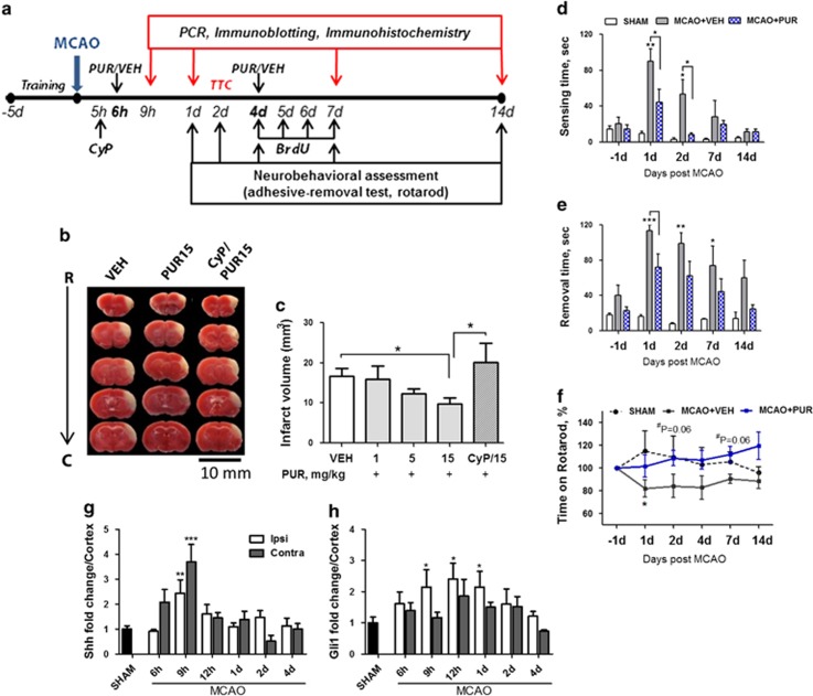
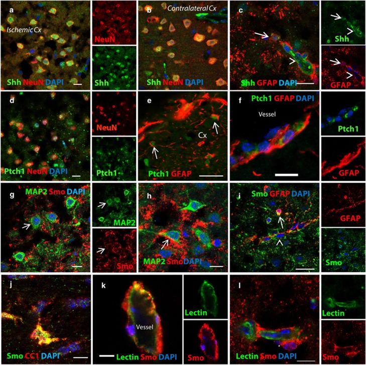
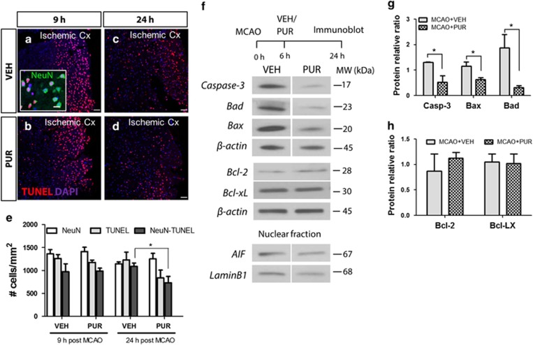
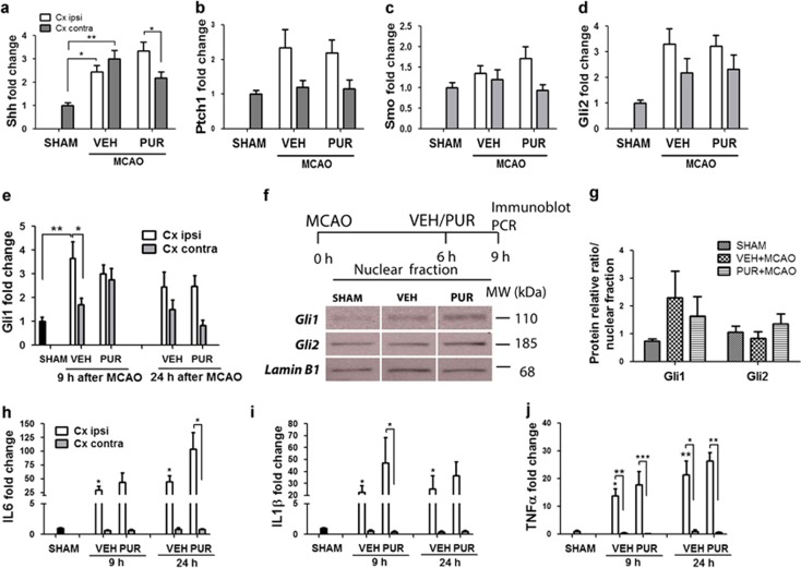
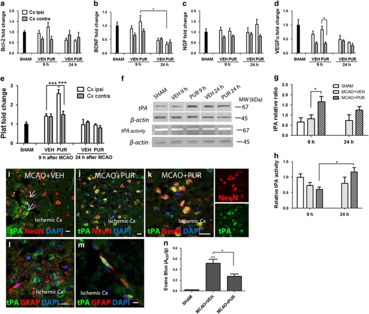
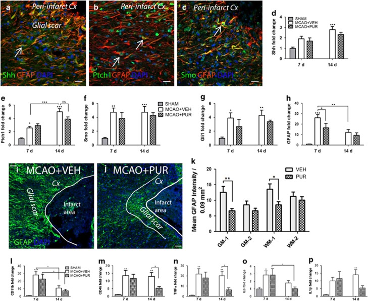
Ischemic stroke occurs as a result of blood supply interruption to the brain causing tissue degeneration, patient disabilities or death. Currently, treatment of ischemic stroke is limited to thrombolytic therapy with a narrow time window of administration. The sonic hedgehog (Shh) signaling pathway has a fundamental role in the central nervous system development, but its impact on neural cell survival and tissue regeneration/repair after ischemic stroke has not been well investigated. Here we report the neuroprotective properties of a small-molecule agonist of the Shh co-receptor Smoothened, purmorphamine (PUR), in the middle cerebral artery occlusion model of ischemic stroke. We found that intravenous administration of PUR at 6 h after injury was neuroprotective and restored neurological deficit after stroke. PUR promoted a transient upregulation of tissue-type plasminogen activator in injured neurons, which was associated with a reduction of apoptotic cell death in the ischemic cortex. We also observed a decrease in blood-brain barrier permeability after PUR treatment. At 14 d postinjury, attenuation of inflammation and reactive astrogliosis was found in PUR-treated animals. PUR increased the number of newly generated neurons in the peri-infarct and infarct area and promoted neovascularization in the ischemic zone. Notably, PUR treatment did not significantly alter the ischemia-induced level of Gli1, a Shh target gene of tumorigenic potential. Thus our study reports a novel pharmacological approach for postischemic treatment using a small-molecule Shh agonist, providing new insights into hedgehog signaling-mediated mechanisms of neuroprotection and regeneration after stroke.


Figures

Figure 1
Figure 1
PUR is neuroprotective in a mouse model of ischemic stroke. (a) Experimental design. PUR or VEH was administered i.v. at 6 h and 4 d after MCAO. Shh antagonist CyP (10 mg/kg) was injected i.v. 40 min before PUR administration. (b) Infarct area detected by TTC staining in animals treated with VEH, PUR (15 mg/kg) or CyP and PUR (15 mg/kg). (c) Quantitative analysis of TTC (n=8–10). Data are the means±S.E.M.; *P<0.05. Time to (d) sense and (e) remove adhesive from the contralateral front pawn in the adhesive-removal test (n=8–9). (f) Time on rotarod shown as the percentage to the time recorded on the last day of training (n=8–10; *P<0.05 compared with SHAM; #VEH versus PUR). mRNA levels of (g) Shh and (h) Gli1 in the cortex after MCAO (n=3–6; *P<0.05; **P<0.01, ***P<0.001)
Figure 2
Figure 2
Shh signaling is upregulated in the ischemic cortex at 9 h after MCAO. Neurons (a) in the ischemic area and (b) in the contralateral cortex express Shh. (c) Some GFAP+ astrocytes (arrow) and endothelial cells (arrow head) express Shh at 9 h after stroke. (d) Neurons in the infarct area express Shh receptor Ptch1. Ptch1+ astrocytes present in the (e) ischemic parenchyma and (f) perivascular units. (g and h) Neurons in the ischemic area express Shh co-receptor Smo. (i) Smo+ parenchymal (arrow) and perivascular (arrow head) astrocytes and (j) oligodendrocytes in the ischemic cortex. Vascular endothelial cells expressing (k) Smo and (l) Smo-negative endothelial cells in the ischemic area. Scale bar, 50 μm for e and i; 20 μm for (c, d, fh, k and l) 10 μm for (a, b and j). Cx, cortex
Figure 3
Figure 3
Effect of PUR on Shh signaling and inflammation. Level of mRNA coding for (a) Shh, (b) Ptch1, (c) Smo and (d) Gli2 in SHAM- (n=6), VEH- (n=11) and PUR-treated animals (n=11) at 9 h after MCAO. (e) Gli1 mRNA level at 9 and 24 h (n=6) after stroke. (f) Immunoblot analysis of Gli1 and Gli2 in the nuclear protein fraction isolated from ischemic cortex at 9 h after stroke (n=4–5). (g) Quantification of protein level normalized to loading control Lamin B1 (n=5). Bands cropped from the same blot are separated by a vertical line. mRNA expression level of pro-inflammatory cytokines (h) IL6, (i) IL1β and (j) TNFα detected at 9 (n=11) and 24 h (n=6) after MCAO in VEH- and PUR-treated animals. Data are the means±S.E.M.; *P<0.05; **P<0.01; ***P<0.001
Figure 4
Figure 4
PUR reduces apoptotic cell death in the ischemic cortex after MCAO. Apoptotic cells in the ischemic cortex of (a and c) VEH- and (b and d) PUR-treated animals at 9 and 24 h after MCAO (representative image of n=4–5; scale bar, 50 μm). Apoptotic neurons (insert in panel (a); scale bar, 20 μm). Cx, cortex. (e) Quantitative analysis of apoptotic cells in the ischemic cortex (n=4–5). Data are the means±S.E.M.; *P<0.05. (f) Representative blots for cleaved caspase-3, Bad, Bax, Bcl-2 and Bcl-xL in the cytoplasmic and AIF in the nuclear protein fraction. Bands cropped from the same blot are separated by a vertical line. (g and h) Quantification of protein levels relative to loading control β-actin. Data are the means±S.E.M.; n=6; *P<0.05
Figure 5
Figure 5
PUR increases the expression of tPA in ischemic neurons early after treatment. Levels of mRNA coding for (a) Bcl-2, (b) brain-derived neurotrophic factor (BDNF), (c) nerve growth factor (NGF), (d) vascular endothelial growth factor (VEGF) and (e) Plat at 9 (n=6–11) and 24 h (n=6) after MCAO. (f) Immunoblot analysis of tPA protein level and enzymatic activity after stroke. Quantification of (g) tPA protein level and (h) tPA activity normalized to β-actin (n=6). Bands cropped from the same blot are separated by a vertical line. Data are the means±S.E.M.; *P<0.05; **P<0.01; ***P<0.001. (i) tPA+ neurons in the ischemic cortex at 9 h after MCAO in VEH- (arrows) and (j and k) PUR-treated animals (representative images of n=4). (l) Parenchymal astrocytes in the ischemic cortex did not express tPA at 9 h after stroke (m) while some perivascular astrocytes were tPA+. Scale bar, 20 μm for I-L; 10 μm for M. Cx, cortex. (n) BBB permeability at 10 h after MCAO. Data are the means of n=5±S.E.M.; *P<0.05; ***P<0.001
Figure 6
Figure 6
PUR reduces astrogliosis and inflammation at a later time point after stroke. Reactive astrocytes in the glial scar express (a) Shh, (b) Ptch1 and (c) Smo. Representative images of n=8–10. mRNA expression levels of (d) Shh, (e) Ptch1, (f) Smo, (g) Gli1 and (h) GFAP in the ischemic cortex at 7 and 14 d after stroke (n=6–7). Expression of GFAP in the glial scar at 14 d after MCAO in (i) VEH- and (j) PUR-treated animals. Representative images of n=8–10. (k) Mean GFAP intensity in the glial scar in areas corresponding to grey (GM1) or white (WM1) matter in the peri-infarct zone and areas located at 300 μm from the border to the ischemic zone (GM2, WM2; n=8–10). Levels of mRNA coding for (l) CD11b, (m) CD45, (n) TNFα, (o) IL6 and (p) IL1β at 7 and 14 d after stroke (n=6–7). Data are the mean±S.E.M.; *P<0.05; **P<0.01; ***P<0.001. Scale bar, 50 μm for ac, i and j. Cx, cortex; NS, not significant
Figure 7
Figure 7
PUR promotes regeneration after stroke. Expression of GAP43 in the ischemic zone of (a) VEH- and (b) PUR-treated animals at 14 d after stroke. GAP43+ cells with neuroblast morphology (arrows). (c) DCX+ (arrow heads) and GAP43/DCX+ (arrows) cells in the ischemic cortex of PUR-treated animals. (d) Quantitative analysis of GAP43, DCX and GAP43/DCX+ cells. Cells were counted in eight 300 × 300 μm2 squares in the ischemic area. Data are the mean±S.E.M. of n=8–10; **P<0.01. (e) DCX+ cells form clusters (arrow) and chains (arrowhead) of migrating neuroblasts in the CC. (f) DCX+ cells (arrowhead) found to detach and migrate from SVZ toward the ischemic area. (g) DCX+ cells migrate along blood vessels. (h) Immunoblot for GFAP, GAP43, synaptophysin and β-actin at 14 d after MCAO. Representative blots of n=4. (i) Quantification of protein levels relative to loading control β-actin. Data are the means of n=4±S.E.M.; *P<0.05. Expression of synaptophysin in the peri-infarct area of (j) VEH- and (k) PUR-treated animals. (l) Newly generated endothelial cells found in the penumbra (arrow). (m) Neovascularization in the infarct area. Bromodeoxyuridine (BrdU)/CD31 labels endothelial cells generated between 4 and 7 d after MCAO (arrows). (n) Newly generated endothelial cells as part of large size vessels in the infarct area. (o) Quantitative analysis of vascular density (CD31+ vessels) and newly generated (BrdU/CD31+) endothelial cells found in the penumbra and ischemic core at 14 d after stroke. Vascular density and endothelial cell numbers were analyzed in two 300 × 300 μm2 squares placed in the peri-infarct or infarct cortex. Data are the means of n=5–7±S.E.M. (p) Quantitative analysis of BrdU+, Olig2+ and BrdU/Olig2+ cells. Cells were counted in three 300 × 300 μm2 squares in the peri-infarct area of VEH- and PUR-treated animals. Data are the mean±S.E.M.; n=5–7. Scale bar, 50 μm for (ac, e, f, j, k and m) 20 μm for (g and l) N. CP, caudate putamen; Cx, cortex; LV, lateral ventricle

References

    1. 1Shamy MC, Jaigobin CS. The complexities of acute stroke decision-making: a survey of neurologists. Neurology 2013; 81: 1130–1133. - PubMed
    1. 2Traiffort E, Angot E, Ruat M. Sonic Hedgehog signaling in the mammalian brain. J Neurochem 2010; 113: 576–590. - PubMed
    1. 3Charytoniuk D, Porcel B, Rodriguez Gomez J, Faure H, Ruat M, Traiffort E. Sonic Hedgehog signalling in the developing and adult brain. J Physiol Paris 2002; 96: 9–16. - PubMed
    1. 4Hynes M, Porter JA, Chiang C, Chang D, Tessier-Lavigne M, Beachy PA et al. Induction of midbrain dopaminergic neurons by Sonic hedgehog. Neuron 1995; 15: 35–44. - PubMed
    1. 5Lu QR, Yuk D, Alberta JA, Zhu Z, Pawlitzky I, Chan J et al. Sonic hedgehog--regulated oligodendrocyte lineage genes encoding bHLH proteins in the mammalian central nervous system. Neuron 2000; 25: 317–329. - PubMed

Publication types

MeSH terms