

Does optical microangiography provide accurate imaging of capillary vessels?: validation using multiphoton microscopy
- PMID: 25341071
- PMCID: PMC4206785
- DOI: 10.1117/1.JBO.19.10.106011
Does optical microangiography provide accurate imaging of capillary vessels?: validation using multiphoton microscopy
Abstract
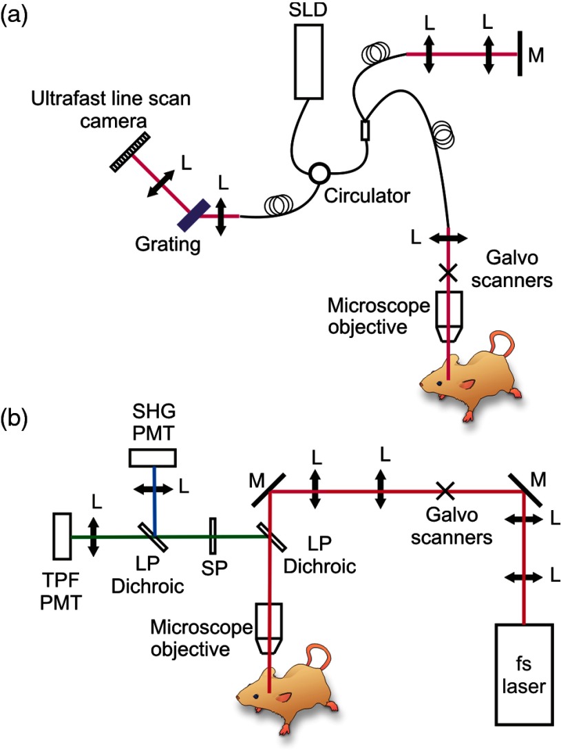
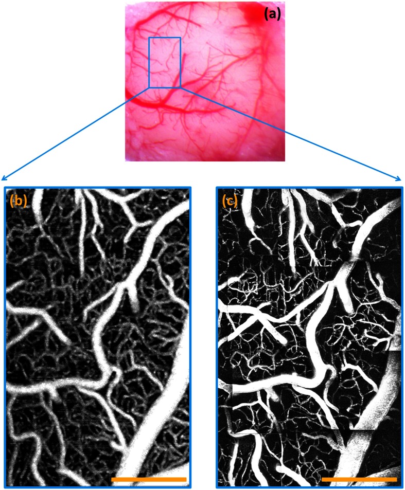
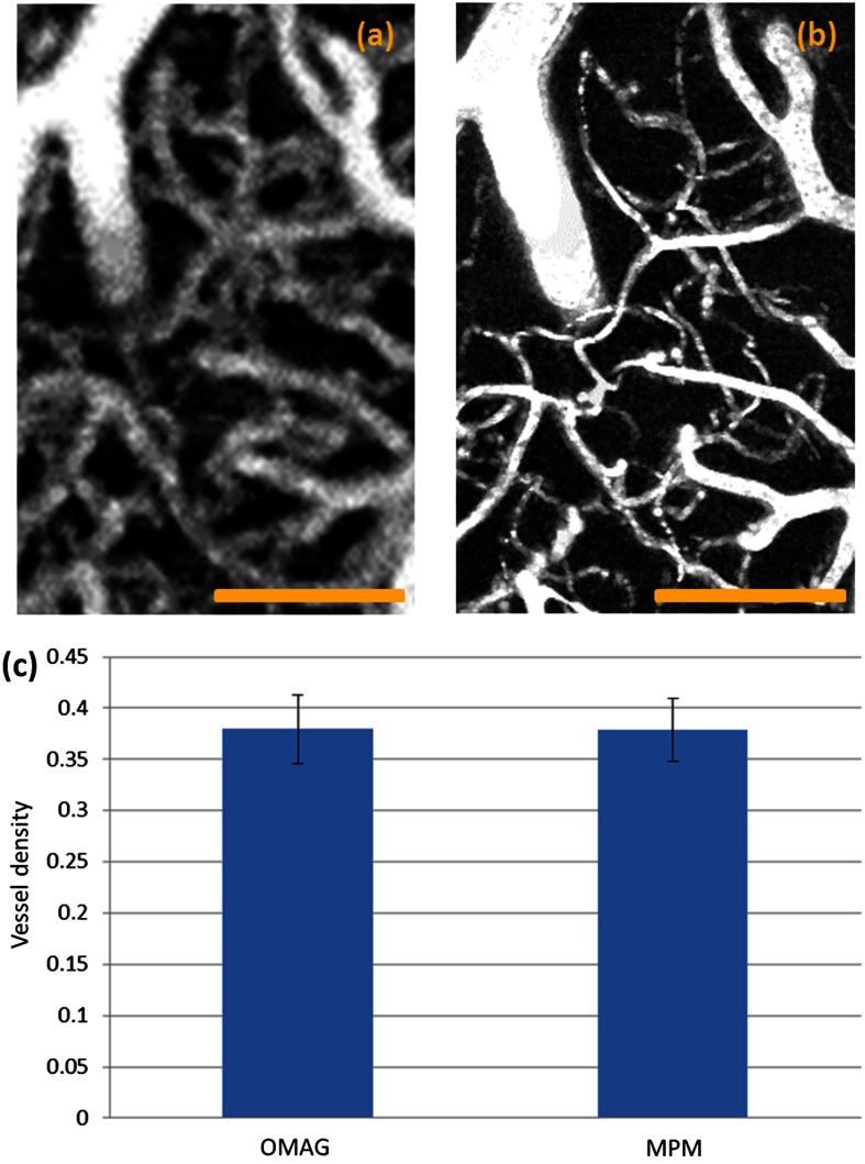
Optical microangiography (OMAG) has been extensively utilized to study three-dimensional tissue vasculature in vivo. However, with the limited image resolution (∼10 μm ) of the commonly used systems, some concerns were raised: (1) whether OMAG is capable of providing the imaging of capillary vessels that are of an average diameter of ∼6 μm ; (2) if yes, whether OMAG can provide meaningful quantification of vascular density within the scanned tissue volume. Multiphoton microscopy (MPM) is capable of depth-resolved high-resolution (∼1 μm ) imaging of biological tissue structures. With externally labeled plasma, the vascular network including single capillaries can be well visualized. We compare the vascular images of in vivo mouse brain acquired by both OMAG and MPM systems. We found that within the penetration depth range of the MPM system, OMAG is able to accurately visualize blood vessels including capillaries. Although the resolution of OMAG may not be able to 100% resolve two closely packed tiny capillaries in tissue, it is still capable of visualizing most of the capillaries because there are interstitial tissue spaces between them. We believe our validation results reinforce the application of OMAG in microvasculature-related studies.
Figures
References
-
- Hunt Thomas K., Wound Healing and Wound Infection: Theory and Surgical Practice, Appleton-Century-Crofts, New York: (1980).
-
- Clark R. A., The Molecular and Cellular Biology of Wound Repair, Springer, New York: (1996).
Publication types
MeSH terms
Grants and funding
LinkOut - more resources
Full Text Sources
Other Literature Sources
