. 2014 Nov 5;136(44):15551-8.
doi: 10.1021/ja5055862. Epub 2014 Oct 23.

Genetically encoded optochemical probes for simultaneous fluorescence reporting and light activation of protein function with two-photon excitation

Affiliations

Genetically encoded optochemical probes for simultaneous fluorescence reporting and light activation of protein function with two-photon excitation

Ji Luo et al. J Am Chem Soc. .

Abstract

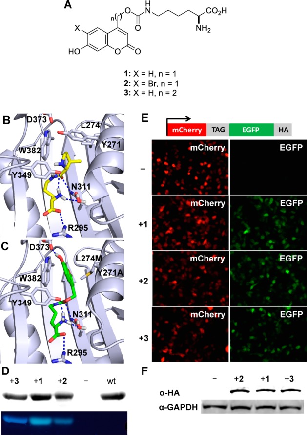
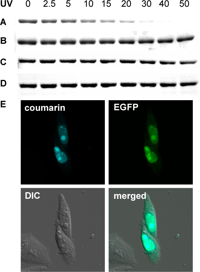
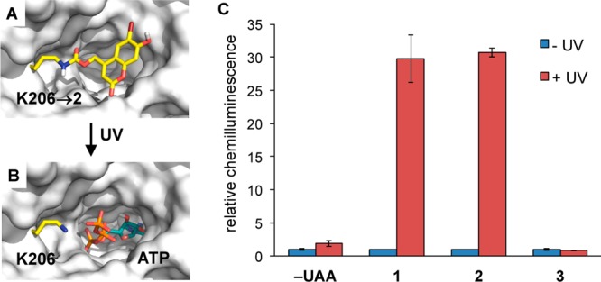
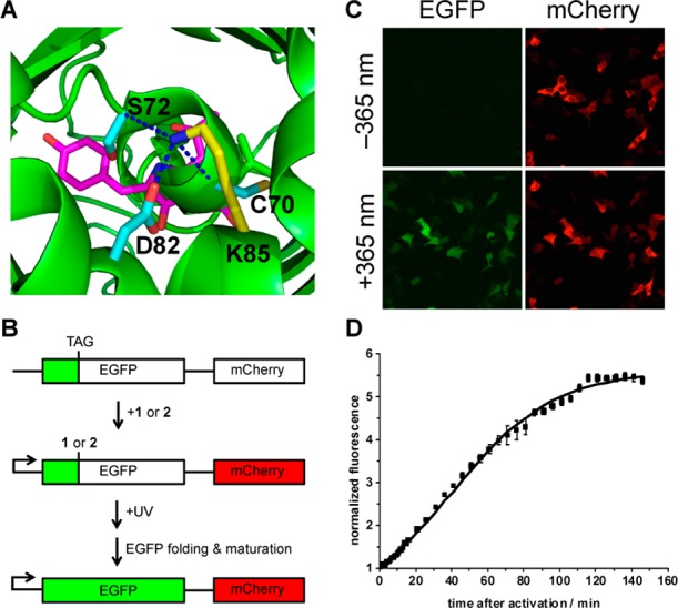
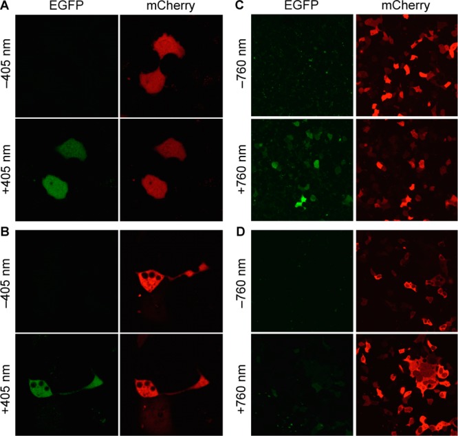
The site-specific incorporation of three new coumarin lysine analogues into proteins was achieved in bacterial and mammalian cells using an engineered pyrrolysyl-tRNA synthetase system. The genetically encoded coumarin lysines were successfully applied as fluorescent cellular probes for protein localization and for the optical activation of protein function. As a proof-of-principle, photoregulation of firefly luciferase was achieved in live cells by caging a key lysine residue, and excellent OFF to ON light-switching ratios were observed. Furthermore, two-photon and single-photon optochemical control of EGFP maturation was demonstrated, enabling the use of different, potentially orthogonal excitation wavelengths (365, 405, and 760 nm) for the sequential activation of protein function in live cells. These results demonstrate that coumarin lysines are a new and valuable class of optical probes that can be used for the investigation and regulation of protein structure, dynamics, function, and localization in live cells. The small size of coumarin, the site-specific incorporation, the application as both a light-activated caging group and as a fluorescent probe, and the broad range of excitation wavelengths are advantageous over other genetically encoded photocontrol systems and provide a precise and multifunctional tool for cellular biology.


Figures

Figure 1
Figure 1
(A) Structures of the genetically encoded coumarin amino acids for fluorescence reporting and light activation of protein function. (B) Crystal structure of PylRS (2Q7H) with the pyrrolysine substrate (yellow) in the active site. (C) Structure of BhcKRS with 1 (green) docked into the active site. Dashed blue lines represent H-bond interactions. (D) SDS-PAGE analysis of sfGFP-Y151TAG containing 13 through incorporation in E. coli. The gel was stained with Coomassie blue (top), and coumarin fluorescence was imaged via excitation at 365 nm (bottom). (E) Fluorescence micrographs of HEK 293T cells expressing the BhcKRS/tRNACUA pair and mCherry-TAG-EGFP-HA in the presence or absence of 13. (F) Western blot analysis of cell lysates using an anti-HA antibody and a GAPDH antibody as a loading control. Full-length protein expression is only observed in the presence of 13, and incorporation efficiency with all three amino acids is similar in mammalian cells.
Figure 2
Figure 2
SDS-PAGE fluorescence analysis shows photodecaging of sfGFP-1 while sfGFP-3 is stable to UV exposure. (A) Loss of coumarin fluorescence after extended sfGFP-1 in-gel decaging for 0–50 min (365 nm, transilluminator). (B) Coomassie staining reveals identical sfGFP-1 protein amounts in all lanes. (C) No loss of coumarin fluorescence is observed, since sfGFP-3 does not decage. (D) Coomassie staining reveals identical sfGFP-3 protein amounts in all lanes. (E) Nuclear colocalization of coumarin and EGFP fluorescence in CHO K1 cells cotransfected with pNLS-TAG-EGFP-HA and the BhcKRS/PylT pair (pBhcKRS-4PylT) in the presence of 1 (0.25 mM). A DIC image and a merged image of all three channels are shown as well.
Figure 3
Figure 3
Engineering of an optochemically controlled Photinus pyralis firefly luciferase through unnatural amino acid mutagenesis. (A) Caging groups at position K206 are blocking access to the binding pocket by luciferin and ATP and are disrupting a required hydrogen bonding network. (B) After decaging, wild-type Fluc is generated and the substrates can now enter the active site. PDB: 2D1S. (C) Bright-Glo luciferase assay of cells that were either kept in the dark or irradiated (365 nm, 4 min). Chemiluminescence units were normalized to the −UAA/–UV control. No enzymatic activity was observed for the caged proteins, and significant increases in luminescence were observed after photolysis of luciferase containing 1 or 2, while the K206 → 3 mutant was permanently deactivated, as expected. Error bars represent standard deviations from three independent experiments.
Figure 4
Figure 4
(A) Location of K85 (yellow) and interactions with D82, C70, and S72 in EGFP. The chromophore is shown in magenta. (B) Schematic of the pEGFP-K85TAG-mCherry construct and its application in light activation studies. (C) Fluorescence imaging of HEK 293T cells expressing EGFP-K85TAG-mCherry, 90 min after irradiation at 365 nm (30 s, DAPI filter, 358–365 nm) in the presence of 1 (Nikon A1R confocal microscope, 20× objective, 2-fold zoom). (D) Normalized EGFP fluorescence as a function of time after 365 nm light activation (error bars represent standard deviations from the measurement of three independent cells, t1/2 = 49 min).
Figure 5
Figure 5
Fluorescence confocal imaging of COS-7 cells expressing EGFP-KTAG-mCherry, before and after irradiation at 405 nm (30 mW diode laser, 20% laser power, 12.6 μs dwell time, 8 cycles) in the presence of 2 (A) or 1 (B) (Zeiss confocal LSM710 microscope, 40× water objective). Similar light-activation experiments before and after irradiation of HEK 293T cells at 760 nm (130 mW, 2 μm/s dwell time, 30 cycles, Olympus Fluoview FV1000 MPE, MaiTai DSBB-OL IR pulsed laser), in the presence of 2 (C) or 1 (D), imaged with a Olympus Fluoview1000, 40× oil objective.

References

    1. Bort G.; Gallavardin T.; Ogden D.; Dalko P. I. Angew. Chem., Int. Ed. 2013, 52, 4526–4537. - PubMed
    1. Krueger A. T.; Imperiali B. ChemBioChem 2013, 14, 788–799. - PubMed
    1. Goncalves M. S. Chem. Rev. 2009, 109, 190–212. - PubMed
    1. Klan P.; Solomek T.; Bochet C. G.; Blanc A.; Givens R.; Rubina M.; Popik V.; Kostikov A.; Wirz J. Chem. Rev. 2013, 113, 119–191. - PMC - PubMed
    1. Brieke C.; Rohrbach F.; Gottschalk A.; Mayer G.; Heckel A. Angew. Chem., Int. Ed. 2012, 51, 8446–8476. - PubMed

Publication types