

Uridine adenosine tetraphosphate is a novel neurogenic P2Y1 receptor activator in the gut
- PMID: 25341729
- PMCID: PMC4226104
- DOI: 10.1073/pnas.1409078111
Uridine adenosine tetraphosphate is a novel neurogenic P2Y1 receptor activator in the gut
Abstract
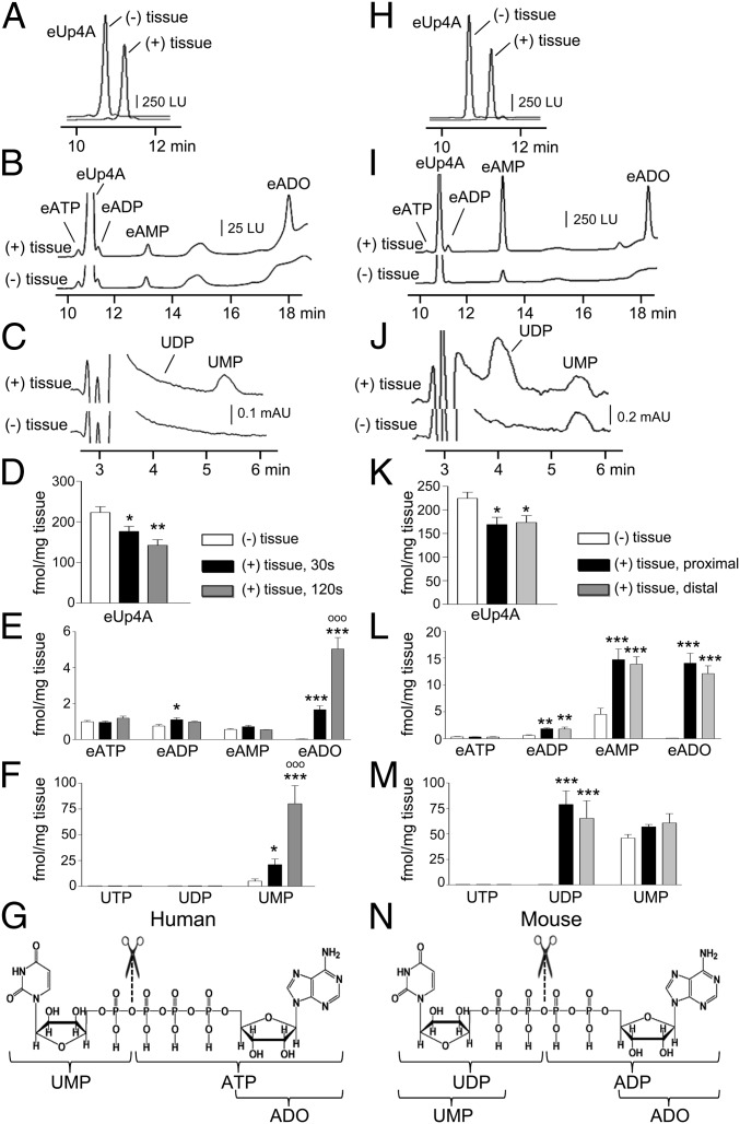
Enteric purinergic motor neurotransmission, acting through P2Y1 receptors (P2Y1R), mediates inhibitory neural control of the intestines. Recent studies have shown that NAD(+) and ADP ribose better meet criteria for enteric inhibitory neurotransmitters in colon than ATP or ADP. Here we report that human and murine colon muscles also release uridine adenosine tetraphosphate (Up4A) spontaneously and upon stimulation of enteric neurons. Release of Up4A was reduced by tetrodotoxin, suggesting that at least a portion of Up4A is of neural origin. Up4A caused relaxation (human and murine colons) and hyperpolarization (murine colon) that was blocked by the P2Y1R antagonist, MRS 2500, and by apamin, an inhibitor of Ca(2+)-activated small-conductance K(+) (SK) channels. Up4A responses were greatly reduced or absent in colons of P2ry1(-/-) mice. Up4A induced P2Y1R-SK-channel-mediated hyperpolarization in isolated PDGFRα(+) cells, which are postjunctional targets for purinergic neurotransmission. Up4A caused MRS 2500-sensitive Ca(2+) transients in human 1321N1 astrocytoma cells expressing human P2Y1R. Up4A was more potent than ATP, ADP, NAD(+), or ADP ribose in colonic muscles. In murine distal colon Up4A elicited transient P2Y1R-mediated relaxation followed by a suramin-sensitive contraction. HPLC analysis of Up4A degradation suggests that exogenous Up4A first forms UMP and ATP in the human colon and UDP and ADP in the murine colon. Adenosine then is generated by extracellular catabolism of ATP and ADP. However, the relaxation and hyperpolarization responses to Up4A are not mediated by its metabolites. This study shows that Up4A is a potent native agonist for P2Y1R and SK-channel activation in human and mouse colon.
Keywords: P2Y1 receptor; Up4A; enteric nervous system; intestine; purinergic signaling.
Conflict of interest statement
The authors declare no conflict of interest.
Figures
Similar articles
-
Platelet-derived growth factor receptor-α-positive cells and not smooth muscle cells mediate purinergic hyperpolarization in murine colonic muscles.Am J Physiol Cell Physiol. 2014 Sep 15;307(6):C561-70. doi: 10.1152/ajpcell.00080.2014. Epub 2014 Jul 23. Am J Physiol Cell Physiol. 2014. PMID: 25055825 Free PMC article.
-
Purinergic inhibitory regulation of murine detrusor muscles mediated by PDGFRα+ interstitial cells.J Physiol. 2014 Mar 15;592(6):1283-93. doi: 10.1113/jphysiol.2013.267989. Epub 2014 Jan 6. J Physiol. 2014. PMID: 24396055 Free PMC article.
-
Diadenosine tetraphosphate activates P2Y1 receptors that cause smooth muscle relaxation in the mouse colon.Eur J Pharmacol. 2019 Jul 15;855:160-166. doi: 10.1016/j.ejphar.2019.05.013. Epub 2019 May 4. Eur J Pharmacol. 2019. PMID: 31063775
-
Progress in understanding of inhibitory purinergic neuromuscular transmission in the gut.Neurogastroenterol Motil. 2013 Mar;25(3):203-7. doi: 10.1111/nmo.12090. Neurogastroenterol Motil. 2013. PMID: 23414428 Free PMC article. Review.
-
Uridine adenosine tetraphosphate and purinergic signaling in cardiovascular system: An update.Pharmacol Res. 2019 Mar;141:32-45. doi: 10.1016/j.phrs.2018.12.009. Epub 2018 Dec 13. Pharmacol Res. 2019. PMID: 30553823 Free PMC article. Review.
Cited by
-
Extracellular metabolism of the enteric inhibitory neurotransmitter β-nicotinamide adenine dinucleotide (β-NAD) in the murine colon.J Physiol. 2020 Oct;598(20):4509-4521. doi: 10.1113/JP280051. Epub 2020 Aug 13. J Physiol. 2020. PMID: 32735345 Free PMC article.
-
Exploring bias in platelet P2Y1 signalling: Host defence versus haemostasis.Br J Pharmacol. 2024 Feb;181(4):580-592. doi: 10.1111/bph.16191. Epub 2023 Aug 2. Br J Pharmacol. 2024. PMID: 37442808 Free PMC article. Review.
-
Mechanisms underlying uridine adenosine tetraphosphate-induced vascular contraction in mouse aorta: Role of thromboxane and purinergic receptors.Vascul Pharmacol. 2015 Oct;73:78-85. doi: 10.1016/j.vph.2015.04.009. Epub 2015 Apr 25. Vascul Pharmacol. 2015. PMID: 25921923 Free PMC article.
-
Transcriptome analysis of mesenchymal stromal cells of the large and small intestinal smooth muscle layers reveals a unique gastrontestinal stromal signature.Biochem Biophys Rep. 2023 Apr 26;34:101478. doi: 10.1016/j.bbrep.2023.101478. eCollection 2023 Jul. Biochem Biophys Rep. 2023. PMID: 37153863 Free PMC article.
-
Nucleotide P2Y1 receptor agonists are in vitro and in vivo prodrugs of A1/A3 adenosine receptor agonists: implications for roles of P2Y1 and A1/A3 receptors in physiology and pathology.Purinergic Signal. 2020 Dec;16(4):543-559. doi: 10.1007/s11302-020-09732-z. Epub 2020 Oct 31. Purinergic Signal. 2020. PMID: 33129204 Free PMC article.
References
-
- Jankowski V, et al. Uridine adenosine tetraphosphate: A novel endothelium- derived vasoconstrictive factor. Nat Med. 2005;11(2):223–227. - PubMed
-
- Jankowski V, et al. Uridine adenosine tetraphosphate acts as an autocrine hormone affecting glomerular filtration rate. J Mol Med (Berl) 2008;86(3):333–340. - PubMed
-
- Jankowski V, et al. Increased uridine adenosine tetraphosphate concentrations in plasma of juvenile hypertensives. Arterioscler Thromb Vasc Biol. 2007;27(8):1776–1781. - PubMed
-
- Matsumoto T, Watanabe S, Kawamura R, Taguchi K, Kobayashi T. Enhanced uridine adenosine tetraphosphate-induced contraction in renal artery from type 2 diabetic Goto-Kakizaki rats due to activated cyclooxygenase/thromboxane receptor axis. Pflugers Arch. 2014;466(2):331–342. - PubMed
Publication types
MeSH terms
Substances
Grants and funding
LinkOut - more resources
Full Text Sources
Other Literature Sources
Molecular Biology Databases
Miscellaneous
