. 2014 Dec;34(12):2601-8.
doi: 10.1161/ATVBAHA.114.304579. Epub 2014 Oct 23.

Loss of β-catenin promotes chondrogenic differentiation of aortic valve interstitial cells

Affiliations

Loss of β-catenin promotes chondrogenic differentiation of aortic valve interstitial cells

Ming Fang et al. Arterioscler Thromb Vasc Biol. 2014 Dec.

Abstract

Objective: The Wnt/β-catenin signaling pathway has been implicated in human heart valve disease and is required for early heart valve formation in mouse and zebrafish. However, the specific functions of Wnt/β-catenin signaling activity in heart valve maturation and maintenance in adults have not been determined previously.

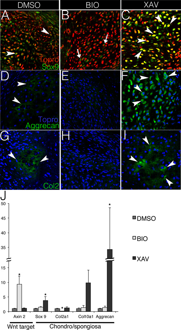
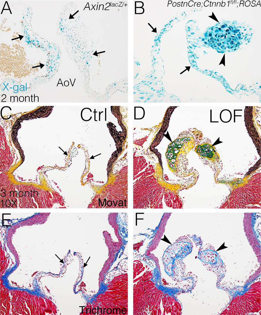
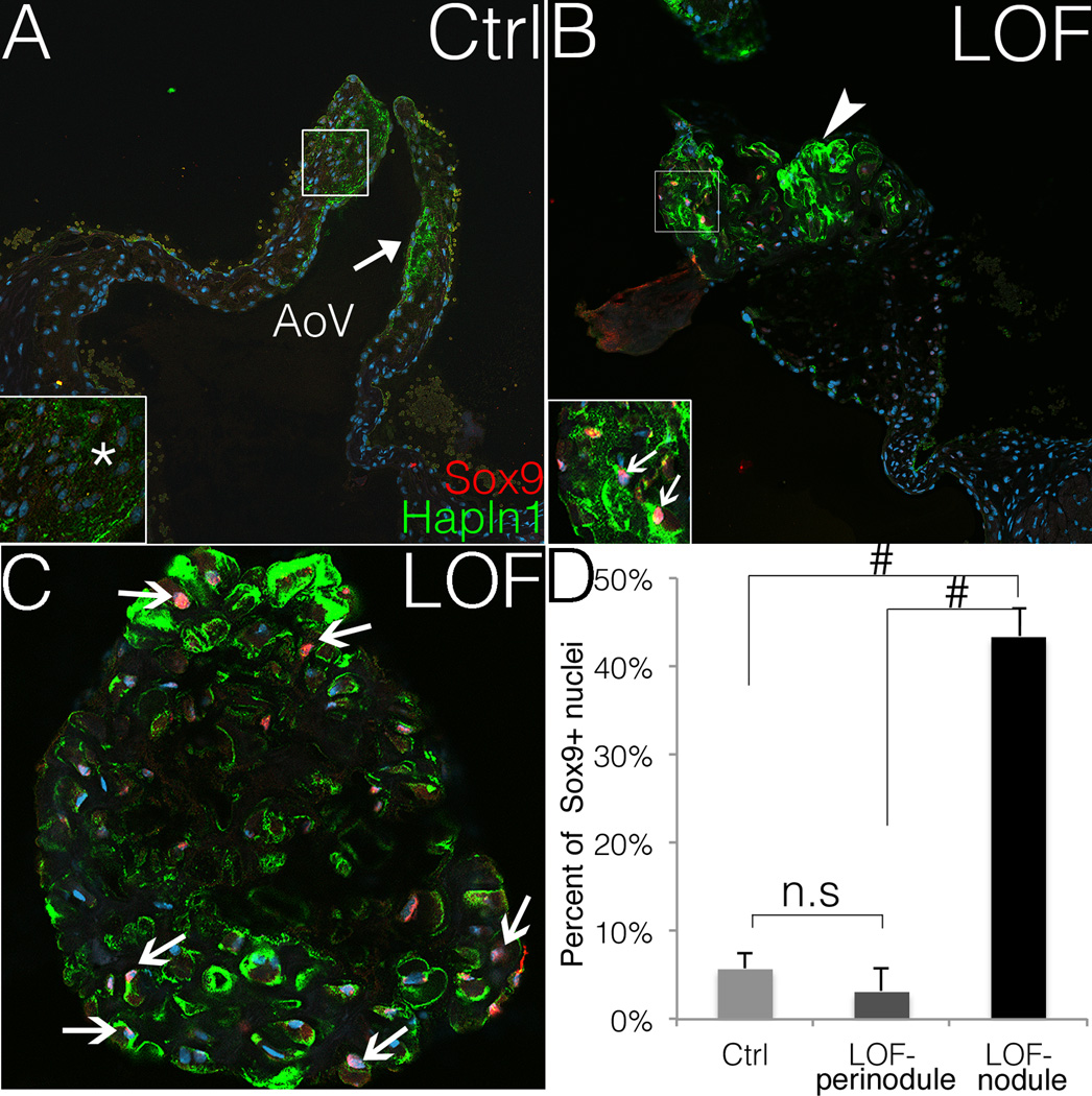
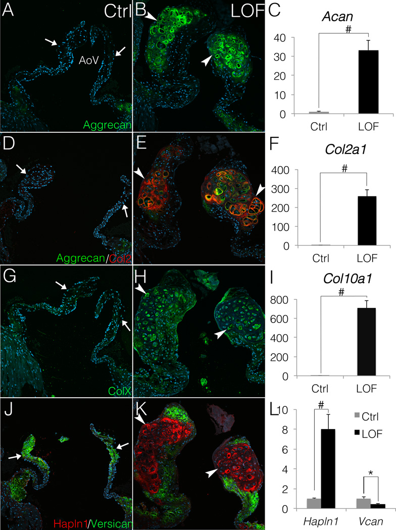
Approach and results: Here, we show that Wnt/β-catenin signaling inhibits Sox9 nuclear localization and proteoglycan expression in cultured chicken embryo aortic valves. Loss of β-catenin in vivo in mice, using Periostin(Postn)Cre-mediated tissue-restricted loss of β-catenin (Ctnnb1) in valvular interstitial cells, leads to the formation of aberrant chondrogenic nodules and induction of chondrogenic gene expression in adult aortic valves. These nodular cells strongly express nuclear Sox9 and Sox9 downstream chondrogenic extracellular matrix genes, including Aggrecan, Col2a1, and Col10a1. Excessive chondrogenic proteoglycan accumulation and disruption of stratified extracellular matrix maintenance in the aortic valve leaflets are characteristics of myxomatous valve disease. Both in vitro and in vivo data demonstrate that the loss of Wnt/β-catenin signaling leads to increased nuclear expression of Sox9 concomitant with induced expression of chondrogenic extracellular matrix proteins.

Conclusions: β-Catenin limits Sox9 nuclear localization and inhibits chondrogenic differentiation during valve development and in adult aortic valve homeostasis.

Keywords: Wnt signaling pathway; aortic valve; chondrogenesis; heart valve disease; proteoglycans.


Figures

Figure 1
Figure 1. Wnt/β-catenin signaling inhibits chondrogenic gene expression in chicken embryo aortic valve organ cultures (aVOC)
A–C. Sox9 expression (green) is shown by immunofluorescent staining in sectioned aVOCs after treatment with DMSO, BIO (Wnt activation) or XAV-939 (Wnt inhibition). Arrowheads indicate cells with positive nuclear Sox9 staining (orange) in A and C, whereas arrows in B indicate cells with cytosolic Sox9 (green). D–F. Aggrecan expression (green) is detected by immunofluorescent staining in treated aVOCs. Arrowheads indicate positive Aggrecan staining in both D and F. G–I. Col2 expression (green, arrowheads) is decreased in BIO-treated aVOCs (H), compared to either DMSO controls (G) or XAV-treated aVOCs (I). Arrowheads indicate positive Col2 staining in both G and I. Nuclei are counterstained with Topro 3, and pseudo-colored in red (A–C), or blue (D–I). Representative images are shown from four aVOCs analyzed for each condition. J. Expression of the Wnt/β-catenin pathway target gene Axin2 and chondrogenic genes Sox9, Col2a1, Col10 and Aggrecan was evaluated by qRT-PCR in cultured aVOC treated with DMSO, BIO or XAV (n=6). Normalized relative gene expression levels were calculated and compared to DMSO controls that were set to 1.0. Statistical significance was determined using paired Student’s t-tests. * indicates p<0.05. Error bars represent SEM.
Figure 2
Figure 2. Wnt/β-catenin signaling is active in adult AoVs and loss of β-catenin leads to formation of proteoglycan-rich hypertrophic cartilage-like nodules in the AoVs of PostnCre;Ctnnb1fl/fl mice
A. Wnt/β-catenin signaling, as indicated by Axin2lacZ reporter activity, is active in the distal tip of a normal aortic valve (AoV) at 2 months-of-age. Arrows indicate cells with β-gal activity (blue) expressed from the Axin2lacZ locus in X-gal stained sections. B. PostnCre is active throughout the leaflets (arrows) of AoV, including the nodule (arrowheads), in PostnCre;Ctnnb1fl/fl;ROSA26 mice at 3 months of age. C, D. AoVs are stained by Movat’s Pentachrome to visualize the ECM distribution and morphology at 3 months. Arrows in C indicate normal valve stratification, whereas arrowheads in D indicate proteoglycan-rich (blue) nodules. E, F. AoVs were stained by Masson’s Trichrome for detection of collagen (blue) at 3 months of age (n=4). Arrows in E indicate normal collagen deposition, whereas arrowheads in F indicate collage expression surrounding the nodules.
Figure 3
Figure 3. The nodular cells PostnCre;Ctnnb1fl/fl AoVs express nuclear Sox9
A, B. Sox9 expression (red) is shown in PostnCre;Ctnnb1fl/fl (LOF) nodular cells but is not detected (asterisk) in Cre-negative control (Ctrl) AoVs. The arrow in A indicates normal expression of Hapln1 (green) in controls. The arrowhead in B indicates the Hapln1-positive (green) LOF nodule, and strong nuclear Sox9 expression (red) in LOF nodular cells (arrows, inset in B). Nuclei are counterstained in blue by Topro3. Staining shown is representative of n=4 specimens analyzed, and a minimum of 2 different slides per sectioned sample was stained. C. Nuclear Sox9 (red) is shown at higher magnification within a Hapln1-positive (green) nodule in a PostnCre;Ctnnb1fl/fl AoV. Arrows indicate nuclear Sox9 positive nodular cells. D. The percent of Sox9 positive nuclei is significantly higher in the LOF nodules than in control AoVs or perinodular regions (Hapln-negative) in LOF AoVs (LOF n=6, control n=8; ANOVA). # indicates p<0.01. n.s indicates not significant (p=0.401). Error bars represent SEM.
Figure 4
Figure 4. PostnCre;Ctnnb1fl/fl nodular cells express chondrocyte markers Aggrecan, Col2 and ColX
A–I, Chondrogenic ECM genes are induced and expressed at high levels in PostnCre;Ctnnb1fl/fl (LOF) nodules compared to Cre-negative controls (Ctrl). Aggrecan (A, B, D & E; green), Col2 (D, E; red) and ColX (G, H; green) expression is shown in control AoVs and LOF nodules. Arrows indicate the lack of endogenous chondrogenic ECM expression in control AoV (A, D, and G), whereas arrowheads indicate induced expression in LOF nodules (B, E, and H). By qRT-PCR (C, F and I), Aggrecan (Acan), Col2a1 and Col10a1 are significantly upregulated in LOF AoV, compared to Cre-negative controls (n=3). J, K. The relative distribution of Versican (green) and Halpn1 (red) expression is shown in LOF and control AoVs. Arrows indicate normal Versican expression and limited expression of Hapln1 in control AoVs (J). Arrowheads indicate markedly reduced Versican expression (green) and increased Hapln1 expression (red) in LOF nodules (K). L. By qRT-PCR, Hapln1 is significantly increased in LOF AoV relative to controls, whereas Versican (Vcan) expression is decreased in LOF AoV compared to controls (n=3). Student’s t-tests were performed to determine the statistical significance of gene expression differences between LOF and control AoVs. # indicates p<0.01, and * indicates p<0.05. All error bars represent SEM.
Figure 5
Figure 5. Fibrosa ECM markers Col1, Col3 and Postn are minimally expressed in the nodules but are present in the perinodular regions
A, B. Col1 expression is shown in PostnCre;Ctnnb1fl/fl (LOF) and Cre-negative control (Ctrl) AoVs. Arrows indicate normal Col1 expression (green) in the valve leaflets, and Hapln1 (red) restricted to the commissure in control AoVs (A). Arrowheads indicate that the Hapln1-positive (red) LOF nodules are Col1 negative, whereas arrows indicate Col1 expression in perinodular regions in LOF AoVs (B). C–F. Similarly, the protein expression (green) of Col3 (C,D) and Postn (E,F) also is decreased in the Hapln1-positive nodules (red) in LOF AoVs (arrowheads, D, F), compared to controls (arrows, C, E). G. By qRT-PCR, Col1a1 and Postn are significantly (n=3; # indicate p<0.01) decreased in LOF AoVs compared to Cre-negative controls as determine by Student’s t-est. However, no significant (n.s.) difference (p=0.438) is detected in Col3a1 expression between controls and LOF mutants. Error bars represent SEM.

Similar articles

Cited by

References

    1. Hinton RB, Yutzey KE. Heart valve structure and function in development and disease. Annu Rev Physiol. 2011;73:29–46. - PMC - PubMed
    1. Hayek E, Gring CN, Griffin BP. Mitral valve prolapse. The Lancet. 2005;365:507–518. - PubMed
    1. Guy TS, Hill AC. Mitral valve prolapse. Annu Rev Med. 2012;63:277–292. - PubMed
    1. Hulin A, Deroanne C, Lambert C, Defraigne J-O, Nusgens B, Radermecker M, Colige A. Emerging pathogenic mechanisms in human myxomatous mitral valve: Lessons from past and novel data. Cardiovasc Pathol. 2013;22:245–250. - PubMed
    1. Lincoln J, Lange AW, Yutzey KE. Hearts and bones: Shared regulatory mechanisms in heart valve, cartilage, tendon, and bone development. Dev Biol. 2006;294:292–302. - PubMed

Publication types

MeSH terms