. 2015 Jan;29(1):336-45.
doi: 10.1096/fj.14-255901. Epub 2014 Oct 23.

Acylcarnitines: potential implications for skeletal muscle insulin resistance

Affiliations

Acylcarnitines: potential implications for skeletal muscle insulin resistance

Céline Aguer et al. FASEB J. 2015 Jan.

Abstract

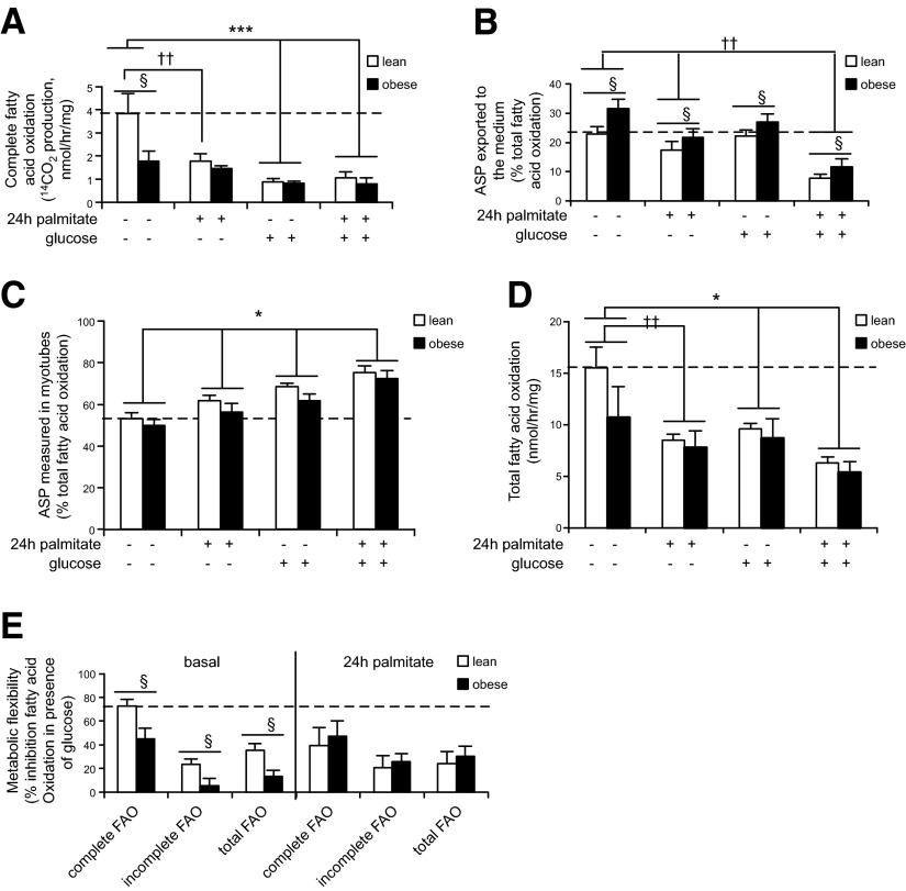
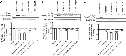
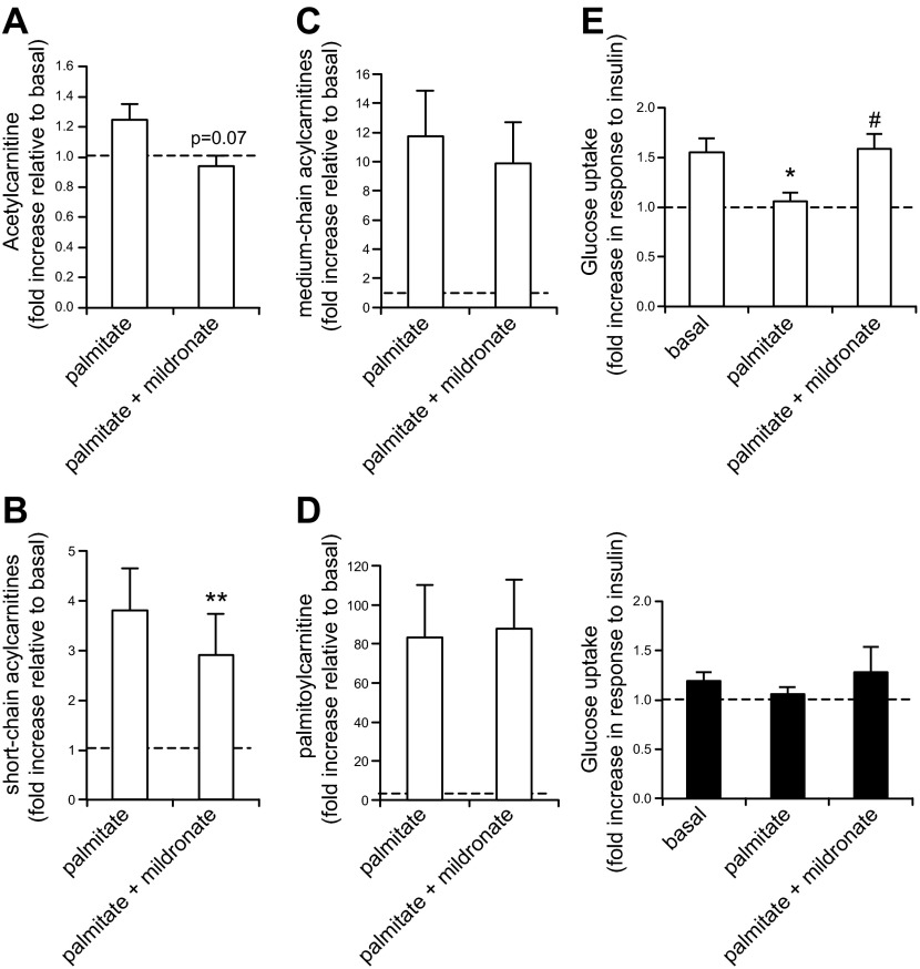
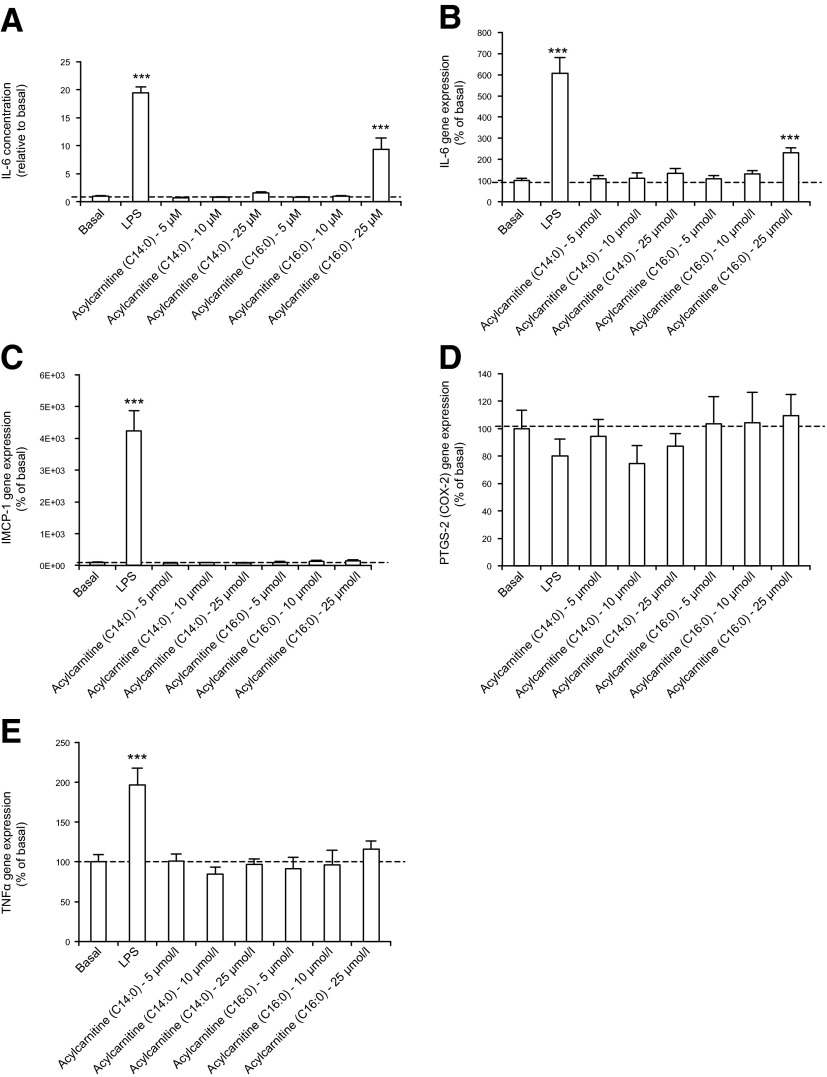
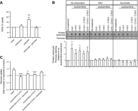
Insulin resistance may be linked to incomplete fatty acid β-oxidation and the subsequent increase in acylcarnitine species in different tissues including skeletal muscle. It is not known if acylcarnitines participate in muscle insulin resistance or simply reflect dysregulated metabolism. The aims of this study were to determine whether acylcarnitines can elicit muscle insulin resistance and to better understand the link between incomplete muscle fatty acid β-oxidation, oxidative stress, inflammation, and insulin-resistance development. Differentiated C2C12, primary mouse, and human myotubes were treated with acylcarnitines (C4:0, C14:0, C16:0) or with palmitate with or without carnitine acyltransferase inhibition by mildronate. Treatment with C4:0, C14:0, and C16:0 acylcarnitines resulted in 20-30% decrease in insulin response at the level of Akt phosphorylation and/or glucose uptake. Mildronate reversed palmitate-induced insulin resistance concomitant with an ∼25% decrease in short-chain acylcarnitine and acetylcarnitine secretion. Although proinflammatory cytokines were not affected under these conditions, oxidative stress was increased by 2-3 times by short- or long-chain acylcarnitines. Acylcarnitine-induced oxidative stress and insulin resistance were reversed by treatment with antioxidants. Results are consistent with the conclusion that incomplete muscle fatty acid β-oxidation causes acylcarnitine accumulation and associated oxidative stress, raising the possibility that these metabolites play a role in muscle insulin resistance.

Keywords: fatty acid β-oxidation; inflammation; mitochondria; myotubes; oxidative stress.


Figures

Figure 1.
Figure 1.
Myotubes derived from obese insulin-resistant subjects release more metabolites associated with incomplete fatty acid β-oxidation and are metabolically inflexible. A) Complete fatty acid β-oxidation (14CO2 production from radiolabeled 1-14C-palmitate) measured in human primary myotubes derived from lean (white bars) and obese (black bars) subjects. §P < 0.05: lean vs. obese. ***P < 0.001: basal vs. glucose. ††P < 0.01: basal vs. palmitate. Group × condition effect: P < 0.05. B) Incomplete fatty acid β-oxidation (14C ASP released in the medium from radiolabeled palmitate) measured in human primary myotubes derived from lean (white bars) and obese (black bars) subjects. §P < 0.05: lean vs. obese. ††P < 0.01: basal vs. palmitate. C) Incomplete fatty acid β-oxidation (14C ASP measured in cell lysate) measured in human primary myotubes derived from lean (white bars) and obese (black bars) subjects. *P < 0.05: treatment effect. D) Total fatty acid β-oxidation (14CO2 + 14C-ASP) measured in human primary myotubes derived from lean (white bars) and obese (black bars) subjects. *P < 0.05: basal vs. glucose. ††P < 0.01, basal vs. palmitate. E) Metabolic flexibility measured as the percentage of fatty acid β-oxidation inhibition in presence of glucose/pyruvate. §P < 0.05: lean vs. obese. A–E) See Materials and Methods for details concerning myotube treatments. n = 5, each measurement done in triplicate. Data are shown as mean ± sem.
Figure 2.
Figure 2.
Acylcarnitine treatments result in insulin resistance at the level of Akt phosphorylation in C2C12 myotubes. A–C) Top panel: representative Western blot of p-Akt (Ser473) expression in C2C12 myotubes. Bottom panel: quantification by density analysis of fold increase in p-Akt (Ser473) relative to basal. C2C12 myotubes were pretreated for 18 h with 750 μmol/L palmitate or 5, 10, or 25 μmol/L (A) C4:0, (B) C14:0, or (C) C16:0 acylcarnitine. p-Akt was calculated relative to total Akt 1/2, and results are expressed as fold of insulin-stimulation of p-Akt in vehicle controls. Akt 1/2 is shown as a loading control. *P < 0.05, **P < 0.01, ***P < 0.001, treated cells vs. basal. Data are shown as mean ± sem.
Figure 3.
Figure 3.
Moderate carnitine acyltransferase inhibition by mildronate restores palmitate-induced insulin resistance in human primary myotubes. A–D) Acylcarnitine measured in the medium of human primary myotubes pretreated with 600 μmol/L palmitate ± 1 mmol/L mildronate for 24 h. n = 3, each individual culture done in duplicate. A) Acetylcarnitine. P = 0.07 palmitate vs. palmitate + mildronate. B) Sum of short-chain acylcarnitines (C4:0 and C6:0 acylcarnitines). **P < 0.01: palmitate vs. palmitate + mildronate. C) Sum of C8:0 to C14:0 acylcarnitines. D) Palmitoyl-carnitine (C16:0). E) Glucose uptake measured in human primary myotubes derived from lean (top panel, white bars) and obese subjects (bottom panel, black bars) pretreated with 600 μmol/L palmitate ± 1 mmol/L mildronate for 24 h. Glucose uptake is presented as the fold increase in response to insulin. *P < 0.05: basal vs. palmitate, #P < 0.05: palmitate vs. palmitate + mildronate. n = 5, each measurement done in triplicate. A–E) See Materials and Methods for details concerning myotube treatments. Data are shown as mean ± sem.
Figure 4.
Figure 4.
Acylcarnitine treatment does not affect C2C12 myotube cytokine secretion at concentrations that elicit insulin resistance. A) IL-6 secretion of C2C12 myotubes. B) IL-6, (C) iMCP1, (D) PTGS-2, (E) TNF-α gene expression in C2C12 myotubes. A–E) See Materials and Methods for details concerning myotube treatments. ***P < 0.001, treated cells vs. basal. Data are shown as mean ± sem.
Figure 5.
Figure 5.
Acylcarnitine treatment of primary myotubes results in increased oxidative stress and insulin resistance, which is partially restored by antioxidant treatment. A) ROS emission measured in mouse primary myotubes with DCFH-DA. **P < 0.01, treated cells vs. basal. n = 4, each measurement done in 5 replicates. B) Top panel: representative dot blot of total protein carbonyl expression in primary myotubes derived from lean subjects. Ponceau staining was used as a loading control. Bottom panel: quantification of protein carbonyls by density analysis. *P < 0.05, treated cells vs. basal. n = 5. C) Glucose uptake measured in primary myotubes derived from lean subjects. Glucose uptake is presented as the fold increase in response to insulin. Basal absolute values (mean; range): 319; 210–447 pmol/min/mg. n = 5, each measurement done in triplicate. A–C) See Materials and Methods for details concerning myotube treatments. Data are shown as mean ± sem.

References

    1. Befroy D. E., Petersen K. F., Dufour S., Mason G. F., de Graaf R. A., Rothman D. L., and Shulman G. I. (2007) Impaired mitochondrial substrate oxidation in muscle of insulin-resistant offspring of type 2 diabetic patients. Diabetes 56, 1376–1381 - PMC - PubMed
    1. Kelley D. E., He J., Menshikova E. V., and Ritov V. B. (2002) Dysfunction of mitochondria in human skeletal muscle in type 2 diabetes. Diabetes 51, 2944–2950 - PubMed
    1. Mogensen M., Sahlin K., Fernström M., Glintborg D., Vind B. F., Beck-Nielsen H., and Højlund K. (2007) Mitochondrial respiration is decreased in skeletal muscle of patients with type 2 diabetes. Diabetes 56, 1592–1599 - PubMed
    1. Petersen K. F., Dufour S., Befroy D., Garcia R., and Shulman G. I. (2004) Impaired mitochondrial activity in the insulin-resistant offspring of patients with type 2 diabetes. N. Engl. J. Med. 350, 664–671 - PMC - PubMed
    1. Muoio D. M., Noland R. C., Kovalik J. P., Seiler S. E., Davies M. N., DeBalsi K. L., Ilkayeva O. R., Stevens R. D., Kheterpal I., Zhang J., Covington, J. D., Bajpeyi, S., Ravussin, E., Kraus, W., Koves, T. R., and Mynatt, R. L. (2012) Muscle-specific deletion of carnitine acetyltransferase compromises glucose tolerance and metabolic flexibility. Cell Metab. 15, 764–777 - PMC - PubMed

Publication types

MeSH terms