. 2014 Dec 5;289(49):33945-57.
doi: 10.1074/jbc.M114.597773. Epub 2014 Oct 23.

Dissection of hexosyl- and sialyltransferase domains in the bifunctional capsule polymerases from Neisseria meningitidis W and Y defines a new sialyltransferase family

Affiliations

Dissection of hexosyl- and sialyltransferase domains in the bifunctional capsule polymerases from Neisseria meningitidis W and Y defines a new sialyltransferase family

Angela Romanow et al. J Biol Chem. .

Abstract

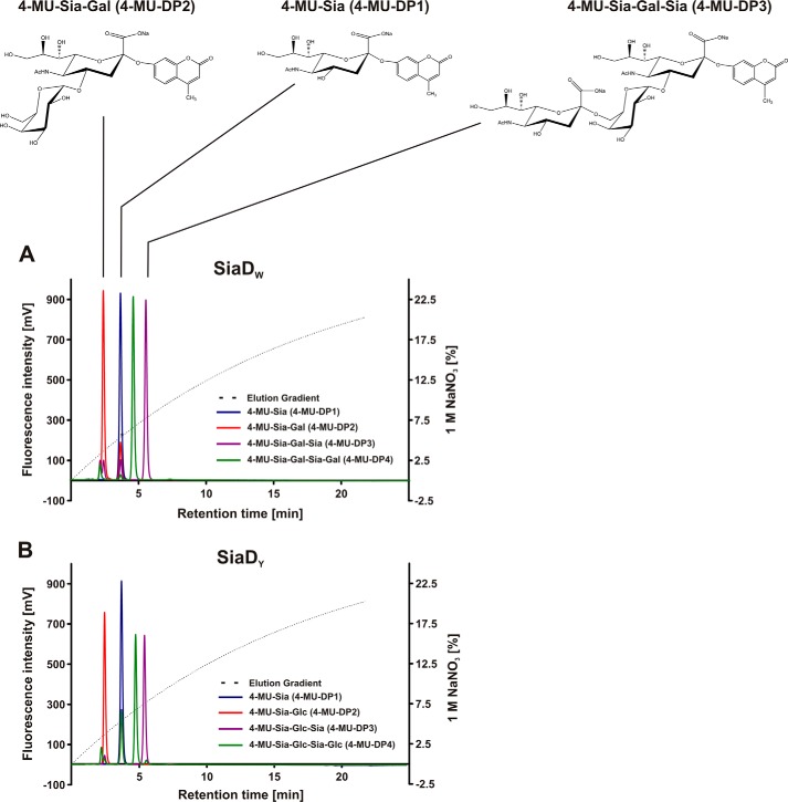
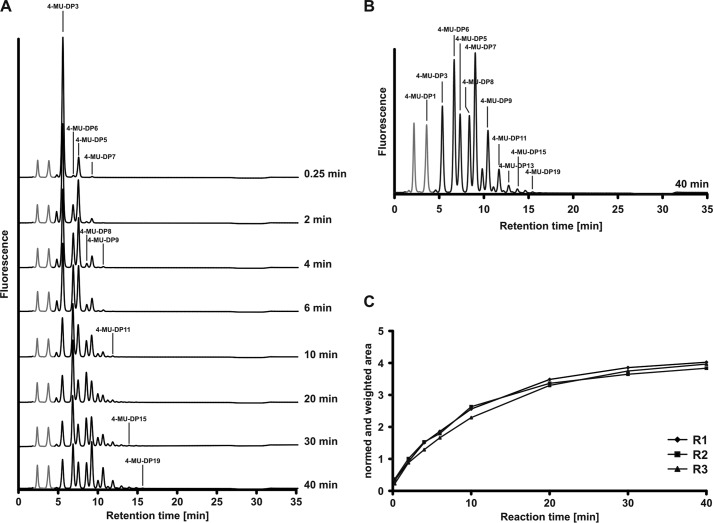
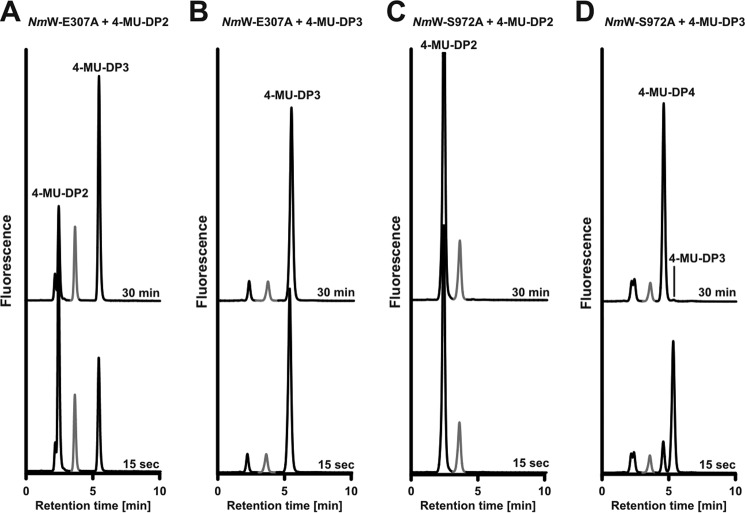
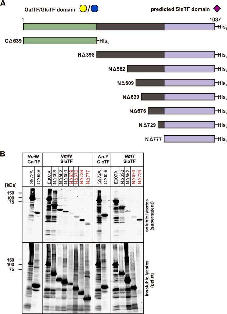
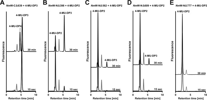
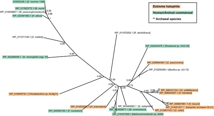
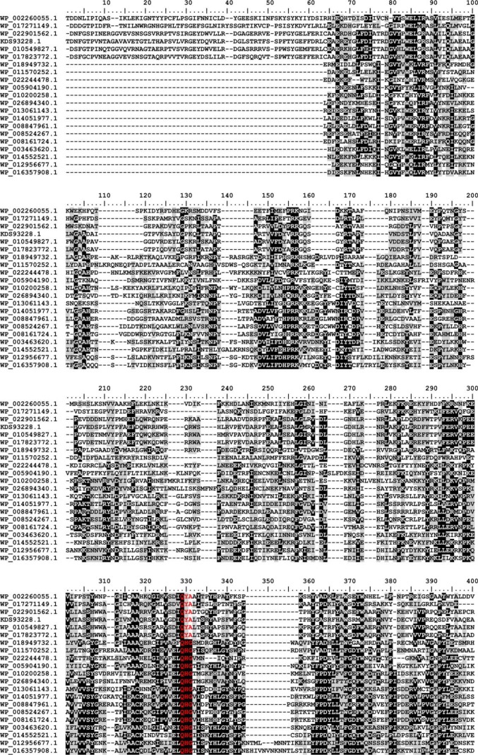
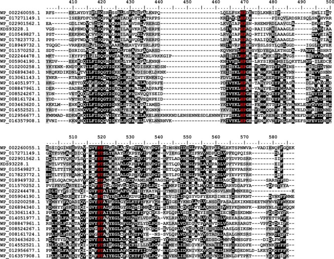
Crucial virulence determinants of disease causing Neisseria meningitidis species are their extracellular polysaccharide capsules. In the serogroups W and Y, these are heteropolymers of the repeating units (→6)-α-d-Gal-(1→4)-α-Neu5Ac-(2→)n in NmW and (→6)-α-d-Glc-(1→4)-α-Neu5Ac-(2→)n in NmY. The capsule polymerases, SiaDW and SiaDY, which synthesize these highly unusual polymers, are composed of two predicted GT-B fold domains separated by a large stretch of amino acids (aa 399-762). We recently showed that residues critical to the hexosyl- and sialyltransferase activity are found in the predicted N-terminal (aa 1-398) and C-terminal (aa 763-1037) GT-B fold domains, respectively. Here we use a mutational approach and synthetic fluorescent substrates to define the boundaries of the hexosyl- and sialyltransferase domains. Our results reveal that the active sialyltransferase domain extends well beyond the predicted C-terminal GT-B domain and defines a new glycosyltransferase family, GT97, in CAZy (Carbohydrate-Active enZYmes Database).

Keywords: Bioinformatics; Capsule Polymerases; Enzyme Catalysis; Fluorescence-based Testing of Glycosyltransferases; Glycosyltransferase; Hexosyltransferases; Neisseria meningitidis Serogroup W and Y; Phylogenetics; Polysaccharide; Sialyltransferases.


Figures

FIGURE 1.
FIGURE 1.
Fluorescently labeled primers for the separate testing of hexosyl- and sialyltransferase activity and chromatographic behavior in anion exchange chromatography. Starting from 4-MU-Sia, the monofunctional active site mutants of SiaDW/SiaDY were used in iterative steps to generate labeled acceptors of defined length. Reaction samples were analyzed on a CarboPac® PA-100 column, and products eluted with a curved NaNO3 gradient as shown. A, structures of the labeled SiaDW glycans (top) and their running behavior on anion exchange chromatography (bottom) are shown. Note that the retention time increases with the addition of a Sia residue and decreases with the addition of Hex, leading to the elution order DP2, DP1, DP4, and then DP3. B, the elution behavior of labeled SiaDY glycans is similar to that of SiaDW. Of note, the chromatograms show that the conversion of 4-MU-DP1 to 4-MU-DP2 was incomplete under the conditions used, whereas all other reactions proceeded to completion.
FIGURE 2.
FIGURE 2.
Time lapse recording of the SiaDW reaction. A, SiaDW was primed with 1 mm 4-MU-Sia-Gal-Sia (4-MU-DP3) in the presence of 2 mm UDP-Gal and 2 mm CMP-Sia. At the indicated time points, product profiles were recorded by HPLC-FD. Gray peaks, 4-MU and 4-MU-DP1, which were left in the priming 4-MU-DP3 pool. Due to the steep increase in acceptor quality from DP2 to DP3, both peaks remained unchanged over the reaction course. B, enlarged display of the HPLC-FD profile obtained at 40 min and labeling of individual DPs. C, reaction progress curves were generated by quantification of each chromatogram and plotting these values against the reaction time point. Three independent experiments (R1–R3) document the high reproducibility of the reaction.
FIGURE 3.
FIGURE 3.
Use of fluorescent primers to monitor the reaction products of single point mutant enzymes. Reaction profiles of the monofunctional mutants NmW-(E307A)-His6 and NmW-(S972A)-His6 were recorded in the presence of both donor sugars using the fluorescent primers as indicated. Reaction samples were taken after 15 s and 30 min. Peaks marked in gray represent 4-MU-DP1 and are unmodified in all reactions. A, NmW-(E307A)-His6 with 4-MU-DP2. The only reaction product formed was 4-MU-DP3. B, NmW-(E307A)-His6 primed with 4-MU-DP3. No product was formed. C, NmW-(S972A)-His6 primed with 4-MU-DP2. No product was formed. D, NmW-(S972A)-His6 primed with 4-MU-DP3. The only product formed was 4-MU-DP4.
FIGURE 4.
FIGURE 4.
The capsule polymerases from NmW and NmY and deletion mutants made thereof. A, schematic illustration of the full-length capsule polymerases NmW/NmY. The GT-B folds predicted to comprise HexTF and SiaTF are highlighted in green and purple, respectively (13). The linker region connecting the GT-B folded domains is shown in black. Mutants made in the course of this project were named according to the introduced deletion. All proteins were expressed with an N-terminal His6 tag. B, Western blot to monitor protein expression. Constructs as indicated were expressed in BL21(DE3), transformed cells were lysed, and lysates were separated into soluble and insoluble fractions. After electrophoretic separation on 10% SDS-PAGE and Western blotting, proteins were displayed with an anti-penta-His antibody. Protein variants that were inactive in the in vitro test system are marked in red.
FIGURE 5.
FIGURE 5.
Activity testing of truncation mutants. Truncation mutant comprising the HexTF (A) and the SiaTF with variant N-terminal extensions (B–E) were assayed in the presence of both donor sugars and a suited fluorescently labeled acceptor as indicated. Reaction samples were taken after 15 s and 30 min. Peaks marked in gray represent 4-MU-DP1. These peaks are unchanged over the reaction course. A, NmW-(CΔ639)-His6 with 4-MU-DP3 catalyzes the transfer of UDP-Gal by creating 4-MU-DP4. B, NmW-(NΔ398)-His6 primed with 4-MU-DP2 catalyzes the transfer of CMP-Sia by producing 4-MU-DP3. C, NmW-(NΔ562)-His6 primed with 4-MU-DP2 catalyzes the transfer of CMP-Sia by creating 4-MU-DP3. D, NmW-(NΔ609)-His6 primed with 4-MU-DP2 catalyzes the transfer of CMP-Sia by producing 4-MU-DP3. E, NmW-(NΔ777)-His6 primed with 4-MU-DP2 does not catalyze the transfer of CMP-Sia.
FIGURE 6.
FIGURE 6.
Phylogenetic tree of family GT97. Phylogenetic analysis was carried out using the PhyML software with bootstrapping (23) using the alignment shown in Fig. 7 as input. Protein accession numbers and species names are indicated, and bootstrapping values are given at the nodes. GT97 family members were only found in the bacterial and archaeal domains of life, and the majority of these species are either human or animal commensals (including several opportunistic pathogens) or extreme halophiles, as indicated.
FIGURE 7.
FIGURE 7.
Multiple sequence alignment of family GT97. Protein sequences were aligned with the MAFFT server (G-INS-I strategy; BLOSUM20 scoring matrix; unalignlevel: 0.4) (21) and visualized using BioEdit (42). High conservation is indicated by white letters on a black background, less conserved regions have black letters on a gray background, and the sialyl-motifs are highlighted in red. Accession numbers correspond to SiaDW and uncharacterized proteins from the following organisms: WP_002260055.1 (aa 563–1037/N. meningitidis serogroup W), WP_017271149.1 (aa 545–999/Sinorhizobium meliloti), WP_022901562.1 (aa 653–1117/Humibacter albus), KDS93228.1 (aa 678–1144/Dermabacter hominis 1368), WP_010549827.1 (aa 651–1116/Brachybacterium paraconglomeratum), WP_017823772.1 (aa 664–1129/Brachybacterium muris), WP_018949732.1 (aa 18–458/Thioalkalivibrio sp. ALMg11), WP_011570252.1 (aa 101–539/Roseobacter denitrificans), WP_022244478.1 (aa 8–439/Roseburia sp. CAG:45), WP_005904190.1 (aa 21–464/Fusobacterium nucleatum), WP_010200258.1 (aa 9–467/Bacillus sp. m3-13), WP_026894340.1 (aa 8–467/Clostridiisalibacter paucivorans), WP_013061143.1 (aa 8–446/S. ruber), WP_014051977.1 (aa 44–483/halophilic archaeon DL31), WP_008847961.1 (aa 33–474/H. kocurii), WP_008524267.1 (aa 8–446/H. tiamatea), WP_008161724.1 (aa 9–445/N. sulfidifaciens), WP_003463620.1 (aa 15–456/Gracilibacillus halophilus), WP_014552521.1 (aa 15–457/Halanaerobium praevalens), WP_012956677.1 (aa 15–497/Methanobrevibacter ruminantium), WP_016357908.1 (aa 13–470/Methanobrevibacter sp. AbM4).
FIGURE 7.
FIGURE 7.
Multiple sequence alignment of family GT97. Protein sequences were aligned with the MAFFT server (G-INS-I strategy; BLOSUM20 scoring matrix; unalignlevel: 0.4) (21) and visualized using BioEdit (42). High conservation is indicated by white letters on a black background, less conserved regions have black letters on a gray background, and the sialyl-motifs are highlighted in red. Accession numbers correspond to SiaDW and uncharacterized proteins from the following organisms: WP_002260055.1 (aa 563–1037/N. meningitidis serogroup W), WP_017271149.1 (aa 545–999/Sinorhizobium meliloti), WP_022901562.1 (aa 653–1117/Humibacter albus), KDS93228.1 (aa 678–1144/Dermabacter hominis 1368), WP_010549827.1 (aa 651–1116/Brachybacterium paraconglomeratum), WP_017823772.1 (aa 664–1129/Brachybacterium muris), WP_018949732.1 (aa 18–458/Thioalkalivibrio sp. ALMg11), WP_011570252.1 (aa 101–539/Roseobacter denitrificans), WP_022244478.1 (aa 8–439/Roseburia sp. CAG:45), WP_005904190.1 (aa 21–464/Fusobacterium nucleatum), WP_010200258.1 (aa 9–467/Bacillus sp. m3-13), WP_026894340.1 (aa 8–467/Clostridiisalibacter paucivorans), WP_013061143.1 (aa 8–446/S. ruber), WP_014051977.1 (aa 44–483/halophilic archaeon DL31), WP_008847961.1 (aa 33–474/H. kocurii), WP_008524267.1 (aa 8–446/H. tiamatea), WP_008161724.1 (aa 9–445/N. sulfidifaciens), WP_003463620.1 (aa 15–456/Gracilibacillus halophilus), WP_014552521.1 (aa 15–457/Halanaerobium praevalens), WP_012956677.1 (aa 15–497/Methanobrevibacter ruminantium), WP_016357908.1 (aa 13–470/Methanobrevibacter sp. AbM4).

References

    1. Willis L. M., Whitfield C. (2013) Structure, biosynthesis, and function of bacterial capsular polysaccharides synthesized by ABC transporter-dependent pathways. Carbohydr. Res. 378, 35–44 - PubMed
    1. Willis L. M., Whitfield C. (2013) KpsC and KpsS are retaining 3-deoxy-d-manno-oct-2-ulosonic acid (Kdo) transferases involved in synthesis of bacterial capsules. Proc. Natl. Acad. Sci. U.S.A. 110, 20753–20758 - PMC - PubMed
    1. Stephens D. S., Greenwood B., Brandtzaeg P. (2007) Epidemic meningitis, meningococcaemia, and Neisseria meningitidis. Lancet 369, 2196–2210 - PubMed
    1. Whitfield C., Roberts I. S. (1999) Structure, assembly and regulation of expression of capsules in Escherichia coli. Mol. Microbiol. 31, 1307–1319 - PubMed
    1. Whitfield C. (2006) Biosynthesis and assembly of capsular polysaccharides in Escherichia coli. Annu. Rev. Biochem. 75, 39–68 - PubMed

Publication types

MeSH terms