. 2014 Oct 24;346(6208):481-485.
doi: 10.1126/science.1257481.

High thermodynamic stability of parametrically designed helical bundles

Affiliations

High thermodynamic stability of parametrically designed helical bundles

Po-Ssu Huang et al. Science. .

Abstract

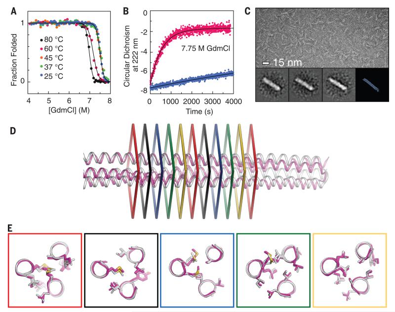
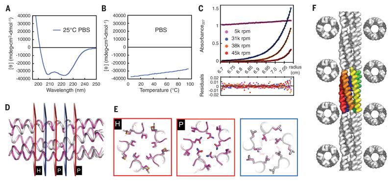
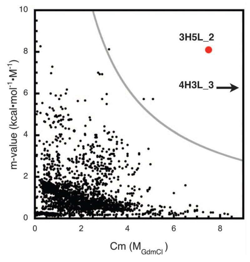
We describe a procedure for designing proteins with backbones produced by varying the parameters in the Crick coiled coil-generating equations. Combinatorial design calculations identify low-energy sequences for alternative helix supercoil arrangements, and the helices in the lowest-energy arrangements are connected by loop building. We design an antiparallel monomeric untwisted three-helix bundle with 80-residue helices, an antiparallel monomeric right-handed four-helix bundle, and a pentameric parallel left-handed five-helix bundle. The designed proteins are extremely stable (extrapolated ΔGfold > 60 kilocalories per mole), and their crystal structures are close to those of the design models with nearly identical core packing between the helices. The approach enables the custom design of hyperstable proteins with fine-tuned geometries for a wide range of applications.


Figures

Fig. 1
Fig. 1. Stability and structure of designed monomeric three-helix bundle 3H5L_2
(A) GdmCl denaturation monitored by CD. At 80°C, the midpoint of the folding transition is ~7 M GdmCl. (B) Kinetics of unfolding in 7.75 M GdmCl at 25°C (blue) and 60°C (red). (C) Negative-stain electron micrographs of 3H5L_2; particle averages are in the inset. The rods are ~12 nm in length, consistent with the 3H5L_2 design model. (D) Superposition of 3H5L_2 crystal structure and design model (RMSD = 3.1 Å over all Cα atoms). Colored rectangles represent the five distinct packing layers in the 18-residue repeat of the structure. (E) Side-chain packing arrangements in each of the five unique layers. Magenta, design model; gray, crystal structure. For each layer, the very similar solutions found by Rosetta in the two central 18-residue repeats are shown.
Fig. 2
Fig. 2. Stability and structure of designed monomeric four-helix bundle 4H3L_3
(A) CD spectra of 4H3L_3 in the presence and absence of GdmCl. (B) Temperature dependence of CD signal at 222 nm in 8 M GdmCl. No unfolding transition is observed at temperatures up to 95°C. (C) DSC of 4H3L_3 in 5 M GdmSCN. An endothermic transition is observed at 97°C (ΔH = 95 kcal/mol). No transition is observed at temperatures up to 130°C in GdmCl or phosphate-buffered saline (PBS) (fig. S5). (D) Superposition of 4H3L_3 crystal structure and design model. At points where the crystal structure deviates from the design model and the helical axis changes direction, peptide backbone carbonyl groups are tipped outward toward the bulk solvent, where they contribute to entrained hydration networks (fig. S6). Colored rectangles indicate the three distinct layers in the 11-residue repeat of the protein. (E) Superposition of 4H3L_3 crystal structure and design model for each of the three unique packing layers for both of the central repeats. Magenta, design model; gray, crystal structure.
Fig. 3
Fig. 3. Stability and structure of designed pentameric five-helix bundle 5H2L_2
(A) CD spectrum and (B) CD-monitored temperature melt of 5H2L_2 (0.2 mg/ml in PBS, pH 7.4). (C) Representative analytical ultra-centrifugation sedimentation-equilibrium curves at four different rotor speeds for 5H2L_2 0.5 mg/ml in PBS, pH 7.4. The data fit (black lines) to a single ideal species in solution corresponding to the pentameric complex of 5H2L_2. (D) Superposition of backbone of crystal structure and design model. The all-atom RMSD between computational model and experimental structure is 0.4 Å. (E) Comparison of side-chain packing in crystal structure (gray) and design model (magenta) at the two unique layers in the 5H2L_2 structure. Two solutions were found for the red layer—a simple aliphatic packing (H) and a polar hydrogen bonding network (P)—and are shown in the two red panels. Both computed solutions were accurately recapitulated in the crystal structure. (F) Packing of the pentamers into straight filaments in the crystal. The colored pentamers occupy one asymmetric unit of the crystal, and the gray pentamers are from adjacent units.
Fig. 4
Fig. 4. High thermodynamic stability of 3H5L_2 and 4H3L_3
X axis, GdmCl denaturation midpoint (Cm); y axis, dependence of folding free energy on GdmCl concentration (m value); black dots, data on previously described proteins from ProTherm database (17); red circle, 3H5L_2; black arrow, lower bound for 4H3L_3 Cm. The free energy of folding in the absence of denaturant is the product of the m-value and the Cm; the curve m-value × Cm = 25 kcal/mol (gray) separates almost all native proteins from the two designs. 4H3L_3 does not denature in GdmCl.

Similar articles

Cited by

References

    1. Boyle AL, Woolfson DN. Chem. Soc. Rev. 2011;40:4295–4306. - PubMed
    1. Crick FHC. Acta Crystallogr. 1953;6:685–689.
    1. Gruber M, Lupas AN. Trends Biochem. Sci. 2003;28:679–685. - PubMed
    1. Hill RB, Raleigh DP, Lombardi A, DeGrado WF. Acc. Chem. Res. 2000;33:745–754. - PMC - PubMed
    1. Harbury PB, Zhang T, Kim PS, Alber T. Science. 1993;262:1401–1407. - PubMed

Publication types

MeSH terms