. 2014 Oct 24;346(6208):1253596.
doi: 10.1126/science.1253596.

Cdc48 and a ubiquitin ligase drive disassembly of the CMG helicase at the end of DNA replication

Affiliations

Cdc48 and a ubiquitin ligase drive disassembly of the CMG helicase at the end of DNA replication

Marija Maric et al. Science. .

Abstract

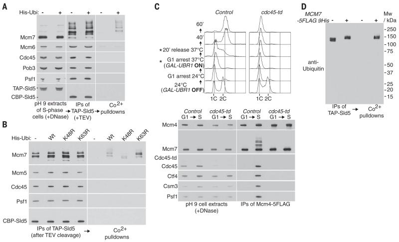
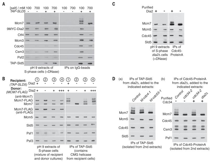
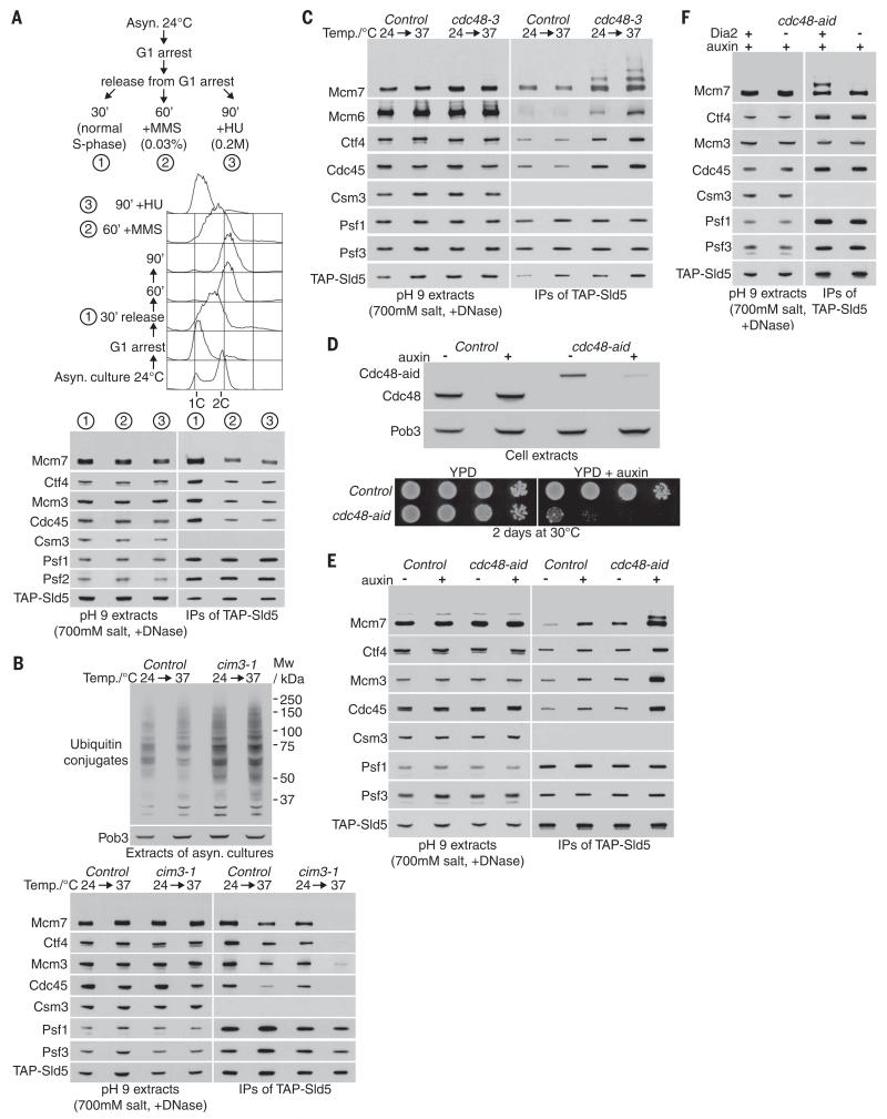
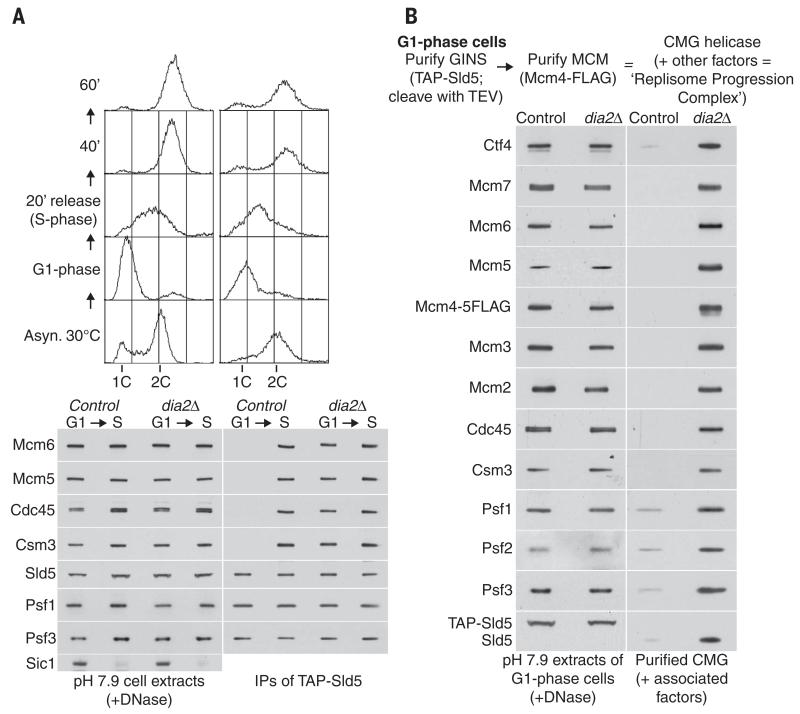
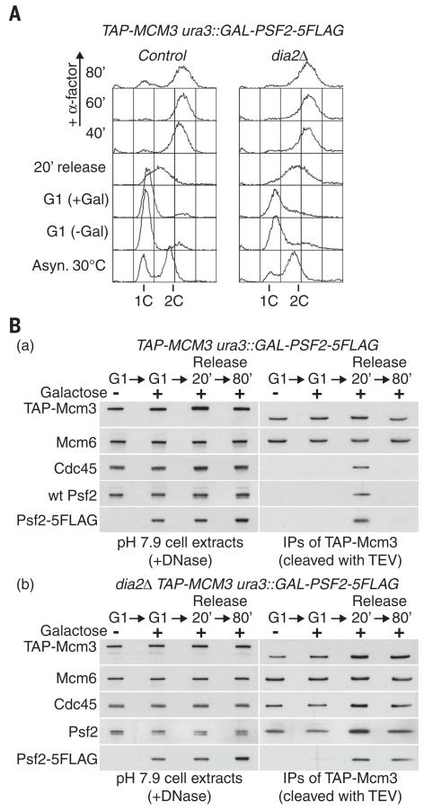
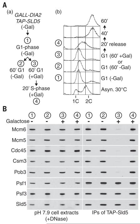
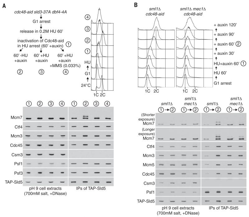
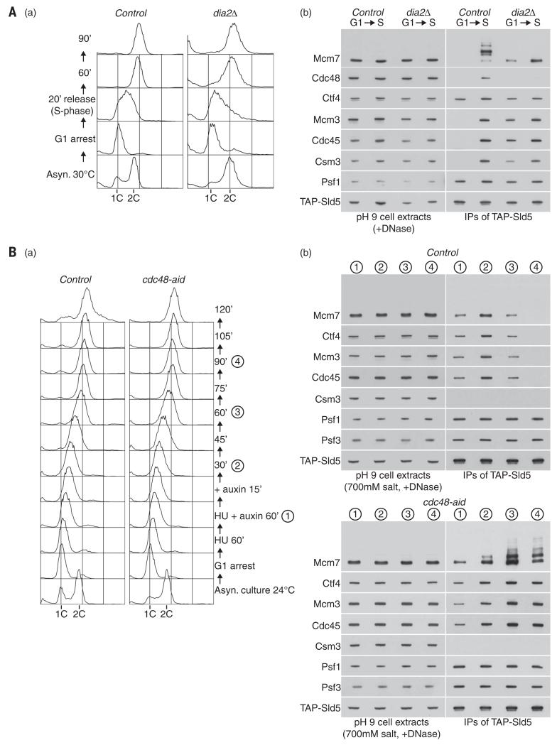
Chromosome replication is initiated by a universal mechanism in eukaryotic cells, involving the assembly and activation at replication origins of the CMG (Cdc45-MCM-GINS) DNA helicase, which is essential for the progression of replication forks. Disassembly of CMG is likely to be a key regulated step at the end of chromosome replication, but the mechanism was unknown until now. Here we show that the ubiquitin ligase known as SCF(Dia2) promotes ubiquitylation of CMG during the final stages of chromosome replication in Saccharomyces cerevisiae. The Cdc48/p97 segregase then associates with ubiquitylated CMG, leading rapidly to helicase disassembly. These findings indicate that the end of chromosome replication in eukaryotes is controlled in a similarly complex fashion to the much-better-characterized initiation step.


Figures

Fig. 1
Fig. 1. The CMG DNA helicase is ubiquitylated on its Mcm7 subunit
(A) Extracts of S-phase cells (YMM22 and YMM23), with or without a plasmid expressing His-tagged ubiquitin, were prepared at pH 9 before digestion of chromosomal DNA and isolation of the GINS component of the CMG helicase, via a TAP tag on the Sld5 subunit. The isolated complexes, representing a mixture of GINS and CMG helicase as part of the replisome progression complex (1), were then released from beads by cleavage with TEV protease before denaturation with 8 M urea (described in the materials and methods section), so that ubiquitylated factors could be isolated specifically on cobalt-coated magnetic beads. IPs, immunoprecipitates. (B) A similar experiment was performed with control cells (YMM22) or cells expressing wild-type (YMM23) ubiquitin, K48R ubiquitin (YMM89), or K63R ubiquitin (YMM90). (C) Control (YSS184) and cdc45-td (YMM74) cells were arrested in G1 phase at 24°C, before inactivation of Cdc45-td and release into S phase for 20 min, as described in the materials and methods. DNA content was measured by flow cytometry (asterisks denote samples that were used to prepare cell extracts). After digestion of chromosomal DNA in cell extracts, the Mcm4 subunit of the Mcm2-7 helicase core was then isolated by immunoprecipitation. This enriched specifically for two forms of Mcm2-7 complex, each of which are bound to chromatin (14): the inactive double hexamer at origins and the active CMG helicase at forks, the latter of which is the substrate for ubiquitylation on its Mcm7 subunit. (D) A similar experiment to that shown in (A) was performed with control cells (YASD375) or cells with tagged Mcm7 (YGDP483). The isolated material was resolved in a 4 to 12% gradient gel, before immunoblotting with anti-ubiquitin antibody (P4D1). Mw, molecular weight. Additional flow cytometry data for the experiments depicted in this figure can be found in fig. S14.
Fig. 2
Fig. 2. SCFDia2 drives in vitro ubiquitylation of CMG in yeast cell extracts
(A) Control (YHM117) and TAP-SLD5 cells (YHM132) were processed as above, except that the extracts were made in the presence of 700 mM salt where indicated. Note that the CMG helicase is still stable in the presence of 700 mM salt but does not associate with other replisome components such as Csm3 or Dia2. (B) To demonstrate that Dia2 promotes the in vitro ubiquitylation of CMG, we synchronized the indicated TAP-SLD5 recipient strains (1, YASD375; 2 to 4, YHM130) and MCM7-5FLAG donor strains (1 to 4, YTM305, YTM306, YTM305, and YTM312, respectively) in S phase at 30°C. Each of the indicated pairs of recipient and donor cultures were then mixed and used to prepare a single-cell extract at pH 9 (see materials and methods). After digestion of chromosomal DNA, the CMG helicase from recipient cells was isolated and monitored by immunoprecipitation of its TAP-Sld5 subunit. (C) dia2Δ CDC45-Protein A cells (YTM415) were synchronized in S phase, as above, and used to prepare a cell extract at pH 9. Purified Dia2 (from YTM532; see materials and methods) was added to the cell extracts as indicated, before immunoprecipitation of Cdc45-Protein A and detection of the associated CMG components by immunoblotting. (D) (a) Material containing CMG complexes was isolated from extracts of S-phase dia2Δ cells (YHM130) by immunoprecipitation of TAP-tagged Sld5 on IgG beads. The beads were then added to pH 9 extracts of control cells (YTM330), td-cdc4-1 cells (YTM445), or td-cdc53-1 cells (YTM444), which had been grown initially at 24°C, before shifting to 37°C for 60 min to inactivate Td-cdc4-1 and Td-cdc53-1 (see materials and methods for details). After reisolation of the beads, the indicated proteins were detected by immunoblotting. (b) Analogous experiments were performed with CMG-containing material that was isolated by immunoprecipitation of Cdc45-Protein from dia2Δ cells (YTM415). In this case, the beads were added to pH 9 extracts of control cells (YTM330), cdc34-2 cells (YTM376), or dia2Δ cells (YMM90), which were then shifted from 24° to 37°C for 60 min. As indicated, the extracts were supplemented with purified Cdc34, which was isolated on IgG beads from an extract of CDC34-TAP cells (YTM396) and then liberated with TEV protease.
Fig. 3
Fig. 3. Dia2-dependent ubiquitylation of CMG in vivo is revealed by inactivation of Cdc48
(A) YASD375 was released into S phase either for 30 min (1), 60 min in the presence of 0.033% methyl methanesulfonate (MMS) (2), or 90 min in the presence of 0.2 M hydroxyurea (HU) (3) and then processed as in Fig. 1. DNA content was measured by flow cytometry. (B) Control (YASD375) and cim3-1 cells (YMM206) were grown at 24°C then incubated for a further hour at 24° or 37°C. The accumulation of ubiquitylated proteins was monitored by immunoblotting with an antibody specific for ubiquitin conjugates (FK2 antibody; upper panels). CMG was monitored as described above, by immunoprecipitation of TAP-tagged Sld5 (lower panels). (C) An analogous experiment to that in (B) was performed with control cells (YASD375) and cdc48-3 (YMM214). (D) The level of Cdc48 in control (YMM256) and cdc48-aid (YMM228) cells was monitored by immunoblotting before and after addition of auxin to the culture medium (upper panels). Serial dilutions of control (YJW15) and cdc48-aid (YMM203) cells were spotted onto the indicated media and incubated for 2 days at 30°C (lower panels). (E) Control (YMM256) and cdc48-aid cells (YMM228) were grown at 24°C, then incubated for 90 min in the presence or absence of auxin, as indicated, before processing as above. (F) Asynchronous cultures of cdc48-aid (YMM228) and cdc48-aid GALL-DIA2 (YMM283) were grown at 30°C in YPD media (see materials and methods for initial growth conditions of cdc48-aid GALL-DIA2), before addition of auxin for 2 hours.
Fig. 4
Fig. 4. CMG is present during the G1 phase of the cell cycle in the absence of Dia2
(A) Control (YASD375) and dia2Δ (YHM130) cells were grown at 30°C, synchronized in G1 phase by addition of mating pheromone, and then released into S phase. Samples were taken at the indicated times (S phase, 20-min sample) and processed for flow cytometry (top panels) or else used to make cell extracts (containing 100 mM salt), before digestion of chromosomal DNA and immunoprecipitation of the TAP-tagged Sld5 subunit of GINS (lower panels). The association of GINS with Cdc45 and Mcm2-7, as well as with other components of the replisome progression complex such as Csm3 and Ctf4 (1), was monitored by immunoblotting of the indicated proteins. (B) Cell extracts were generated as above, from G1 phase control (YAG230-3) and dia2Δ (YTM687) cells. To screen for the presence of the CMG helicase, GINS was isolated as above, before release of the purified material from beads by cleavage with TEV protease and immunoprecipitation of Mcm4 (1). The final material was analyzed by immunoblotting, along with the cell extracts.
Fig. 5
Fig. 5. Disassembly of the CMG helicase at the end of S phase requires Dia2
(A) A plasmid containing the GAL-PSF2-5FLAG construct was integrated at the ura3 locus in control (YTM593) and dia2Δ (YTM592) cells, which were then synchronized at 30°C in G1 phase with mating pheromone, in medium lacking galactose. Cells were held in G1 phase for a further 60 min in the presence of galactose to induce expression of Psf2-5FLAG, before release for the indicated times in medium lacking mating pheromone. Alpha factor mating pheromone was added again from 40 min onward to prevent nuclei from entering into the next round of S phase, and DNA content was measured throughout the experiment by flow cytometry. (B) Cell extracts from the same experiment as depicted in (A) were treated with DNase before immunoprecipitation of TAP-tagged Mcm3. The material was released from beads with TEV protease before immunoblotting, to prevent TAP-Mcm3 from interfering with the similarly sized Mcm6 protein.
Fig. 6
Fig. 6. Dia2 induces disassembly of terminated CMG complexes but does not prevent assembly of active CMG complexes
(A) (a) Cells in which the endogenous DIA2 gene had been placed under the control of the weak and inducible GALL promoter (YTM568) were grown at 30°C in medium lacking galactose (GALL-DIA2 OFF) and then synchronized in G1 phase by addition of mating pheromone (stage 1). The cells were then held for a further 60 min in G1 phase, either in the continued absence of galactose (GALL-DIA2 OFF, stage 2), or in the presence of galactose (GALL-DIA2 ON, stage 3) before subsequent release for 20 min into S phase (GALL-DIA2 ON, stage 4). (b) DNA content was monitored by flow cytometry. (B) Cell extracts were prepared, treated with DNase, and then used to isolate GINS as above, before monitoring of the associated factors by immunoblotting as indicated.
Fig. 7
Fig. 7. The CMG helicase is ubiquitylated in vivo during a late step of DNA replication
(A) cdc48-aid sld3-37A dbf4-4A (YMM309) was synchronized in G1 phase and then released for 60 min into S phase in the presence of 0.2 M hydroxyurea (HU) to allow early and late origins to fire (55). Auxin was then added to inactivate Cdc48-aid (1) before the culture was split and cells were released for 60 min in fresh medium lacking HU (2), held for a further 60 min in the presence of HU (3), or subjected to DNA damage (by addition of 0.033% MMS) in the continued presence of HU (4). The sld3-37A dbf4-4A background was used to allow late origins to fire in the HU arrest, thus ensuring that cells would have a similar number of CMG complexes both before and after release from HU arrest, making easier the comparison of the Mcm7 subunit of CMG in the two cases. DNA content was measured by flow cytometry. (B) A similar experiment was performed with control cells (YMM366, sml1Δ cdc48-aid ADH-TIR1) and mec1Δ (YMM368, mec1Δ sml1Δ cdc48-aid ADH-TIR1) strains. Inactivation of Cdc48-aid in hydroxyurea HU-arrested cells did not cause efficient accumulation of ubiquitylated CMG helicase in either control or mec1Δ cells, consistent with the notion that ubiquitylation only occurs during a late step of DNA replication. When cells were washed into fresh medium lacking hydroxyurea HU, completion of DNA replication in control cells led to the accumulation of ubiquitylated CMG helicase, but this was much less efficient in mec1Δ cells, in which the replisome is stable but is unable to resume remain functional after DNA replication stress (52).
Fig. 8
Fig. 8. Cdc48 is required for disassembly of CMG at the end of chromosome replication
(A) Control (YASD375) and dia2Δ (YHM130) were grown as indicated, and DNA content was measured by flow cytometry (a). GINS was isolated from cell extracts, and the association of the indicated factors (including Cdc48) was monitored by immunoblotting (b). (B) Control (YMM256) and cdc48-aid cells (YMM228) were initially processed as described above for Fig. 7, but were then released from HU arrest for the indicated times in the presence of auxin.

Comment in

Similar articles

Cited by

References

    1. Gambus A, et al. GINS maintains association of Cdc45 with MCM in replisome progression complexes at eukaryotic DNA replication forks. Nat. Cell Biol. 2006;8:358–366. doi:10.1038/ncb1382. - PubMed
    1. Moyer SE, Lewis PW, Botchan MR. Isolation of the Cdc45/Mcm2-7/GINS (CMG) complex, a candidate for the eukaryotic DNA replication fork helicase. Proc. Natl. Acad. Sci. U.S.A. 2006;103:10236–10241. doi:10.1073/ pnas.0602400103. - PMC - PubMed
    1. Li Y, Araki H. Loading and activation of DNA replicative helicases: The key step of initiation of DNA replication. Genes Cells. 2013;18:266–277. doi:10.1111/gtc.12040. - PMC - PubMed
    1. Boos D, Frigola J, Diffley JF. Activation of the replicative DNA helicase: Breaking up is hard to do. Curr. Opin. Cell Biol. 2012;24:423–430. doi:10.1016/j.ceb.2012.01.011. - PubMed
    1. Labib K. How do Cdc7 and cyclin-dependent kinases trigger the initiation of chromosome replication in eukaryotic cells? Genes Dev. 2010;24:1208–1219. doi:10.1101/gad.1933010. - PMC - PubMed

Publication types

MeSH terms

Substances