. 2015 May;21(5):499-508.
doi: 10.1089/ten.TEC.2014.0414. Epub 2015 Mar 31.

Three-dimensional human tissue models that incorporate diabetic foot ulcer-derived fibroblasts mimic in vivo features of chronic wounds

Affiliations

Three-dimensional human tissue models that incorporate diabetic foot ulcer-derived fibroblasts mimic in vivo features of chronic wounds

Anna G Maione et al. Tissue Eng Part C Methods. 2015 May.

Abstract

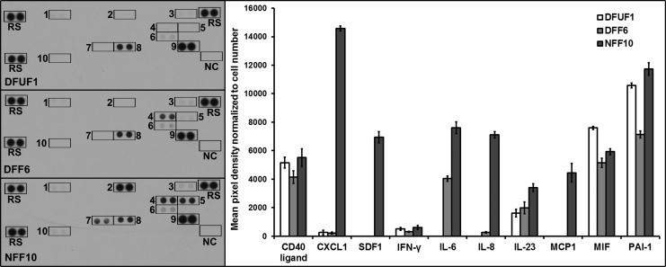
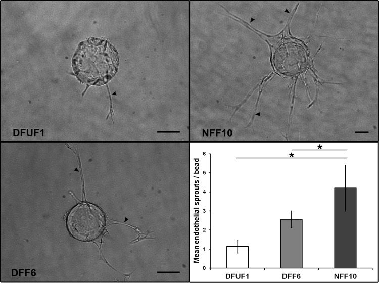
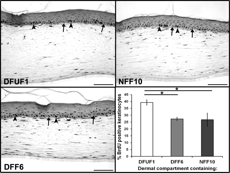
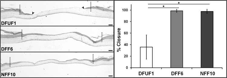
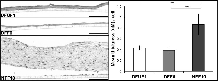
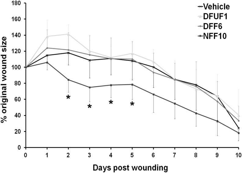
Diabetic foot ulcers (DFU) are a major, debilitating complication of diabetes mellitus. Unfortunately, many DFUs are refractory to existing treatments and frequently lead to amputation. The development of more effective therapies has been hampered by the lack of predictive in vitro methods to investigate the mechanisms underlying impaired healing. To address this need for realistic wound-healing models, we established patient-derived fibroblasts from DFUs and site-matched controls and used them to construct three-dimensional (3D) models of chronic wound healing. Incorporation of DFU-derived fibroblasts into these models accurately recapitulated the following key aspects of chronic ulcers: reduced stimulation of angiogenesis, increased keratinocyte proliferation, decreased re-epithelialization, and impaired extracellular matrix deposition. In addition to reflecting clinical attributes of DFUs, the wound-healing potential of DFU fibroblasts demonstrated in this suite of models correlated with in vivo wound closure in mice. Thus, the reported panel of 3D DFU models provides a more biologically relevant platform for elucidating the cell-cell and cell-matrix-related mechanisms responsible for chronic wound pathogenesis and may improve translation of in vitro findings into efficacious clinical applications.


Figures

<b>FIG. 1.</b>
FIG. 1.
DFUFs and DFFs displayed altered cytokine secretion in vitro. Images of cytokine array membranes used to analyze secretion profiles of DFUF1, DFF6, and NFF10 are shown. Antibody spot locations are labeled: 1=CD40 Ligand, 2=CXCL1, 3=IFN-γ, 4=IL-6, 5=IL-8, 6=IL-23, 7=MCP-1, 8=MIF, 9=PAI-1, 10=SDF-1, RS=Reference spots, and NC=Negative control. Densitometric quantification of array spots normalized to cell number revealed that DFUF1 and DFF6 exhibit altered secretory profiles compared with NFF10 in 2D monolayer culture in vitro. CXCL1, C-X-C motif chemokine 1; DFFs, type II diabetic, nonulcerated foot-derived fibroblasts; DFUFs, type II diabetic foot ulcer-derived fibroblasts; IFN-γ, interferon gamma; IL, interleukin; MCP-1, monocyte chemotactic protein-1; MIF, macrophage migration inhibitory factor; PAI-1, plasminogen activator inhibitor-1; SDF-1, stromal-derived factor 1.
<b>FIG. 2.</b>
FIG. 2.
DFUFs and DFFs promoted less endothelial cell sprouting than NFFs in a 3D angiogenesis model. Representative images of endothelial cell sprouts (arrowheads) off of dextran beads in fibrin gels in coculture with DFUF1, DFF6, or NFF10 are shown. Scale bars, 100 μm. Quantification of mean number of endothelial sprouts per bead±SD (n=4) demonstrated that DFUF1 and DFF6 were less able to promote sprouting compared with NFF10. *p<0.05. 3D, three dimensional; NFFs, nonulcerated foot-derived fibroblasts.
<b>FIG. 3.</b>
FIG. 3.
DFUFs demonstrated increased basal keratinocyte proliferation in a 3D HSE model. HSE tissues were constructed with DFUF1, DFF6, or NFF10 in the dermal compartment. Representative images of tissue sections stained for BrdU incorporation are shown (arrowheads mark examples of BrdU-stained nuclei, and arrows mark examples of BrdU-negative nuclei). Scale bars, 100 μm. Quantification of mean percentage of basal keratinocytes positive for BrdU±SD (n=3) revealed that keratinocytes in HSEs constructed with DFUF1 were more proliferative than those constructed with DFF6 or NFF10. *p<0.05. BrdU, bromo-deoxyuridine; HSE, human skin equivalent.
<b>FIG. 4.</b>
FIG. 4.
DFUFs exhibited impaired support of re-epithelialization in a wounded HSE model. Representative H&E stained sections of 3D re-epithelialization tissue models constructed with DFUF1, DFF6, or NFF10 are shown. The dotted line demarcates the original wound edge. Arrowheads indicate migrating epithelial edge after 72 h on DFUF-supported wounds. Scale bars, 100 μm. Quantification of re-epithelialization expressed as mean percent wound closure±SD (n=3) demonstrated that DFUF1 was less supportive of re-epithelialization compared with either DFF6 or NFF10. *p<0.05. H&E, hematoxylin and eosin.
<b>FIG. 5.</b>
FIG. 5.
DFUFs and DFFs produced and assembled less ECM than NFFs. Representative H&E stained sections of ECM tissues created by DFUF1, DFF6, or NFF10 are shown. Scale bars, 100 μm. Quantification of mean ECM area normalized to tissue length and cell number±SD (n=3). NFF10-produced ECMs were substantially thicker than those produced by either DFUF1 or DFF6. **p<0.01.
<b>FIG. 6.</b>
FIG. 6.
Transplantation of NFFs correlated with improved wound closure in vivo compared with DFUFs and DFFs. Quantification of wound area (expressed as mean percentage of original wound size±SD) of full-thickness excisional wounds in mice after an injection of DFUF1 (n=3), DFF6 (n=5), NFF10 (n=4) in hydrogel, or hydrogel vehicle alone (n=4). NFF10 injection correlated with improved wound closure overall and specifically on days 2–5 postwounding compared with DFUF1, DFF6, or vehicle alone. *p<0.05.

References

    1. Boulton A.J.M., Vileikyte L., Ragnarson-Tennvall G., and Apelqvist J.The global burden of diabetic foot disease. Lancet 366,1719, 2005 - PubMed
    1. Carlson M.W., Alt-Holland A., Egles C., and Garlick J.A.Three-dimensional tissue models of normal and diseased skin. Curr Protoc Cell Biol Editor Board Juan Bonifacino Al Chapter 19, Unit 19.9 2008 - PMC - PubMed
    1. Breslin S., and O'Driscoll L.Three-dimensional cell culture: the missing link in drug discovery. Drug Discov Today 18,240, 2013 - PubMed
    1. Singer A.J., and Clark R.A.Cutaneous wound healing. N Engl J Med 341,738, 1999 - PubMed
    1. Brem H., et al. . Primary cultured fibroblasts derived from patients with chronic wounds: a methodology to produce human cell lines and test putative growth factor therapy such as GMCSF. J Transl Med 6,75, 2008 - PMC - PubMed

Publication types