. 2014 Oct 24;9(10):e111019.
doi: 10.1371/journal.pone.0111019. eCollection 2014.

DDX3X induces primary EGFR-TKI resistance based on intratumor heterogeneity in lung cancer cells harboring EGFR-activating mutations

Affiliations

DDX3X induces primary EGFR-TKI resistance based on intratumor heterogeneity in lung cancer cells harboring EGFR-activating mutations

Koichiro Nozaki et al. PLoS One. .

Abstract

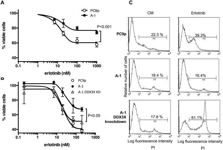
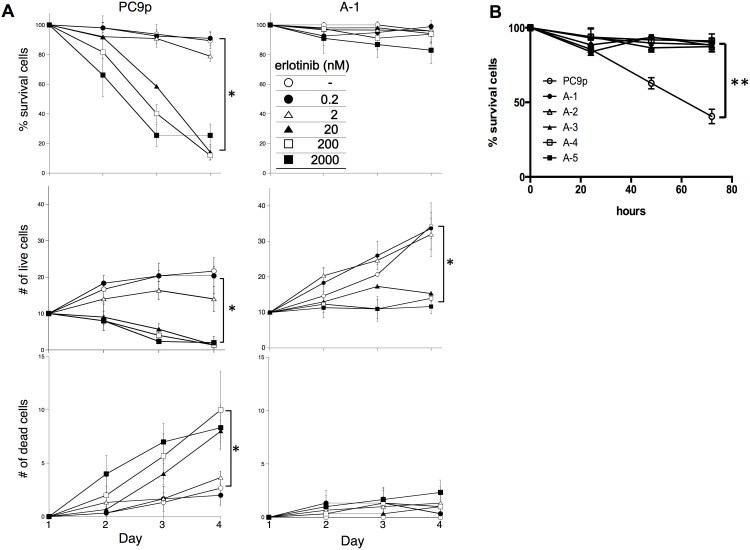
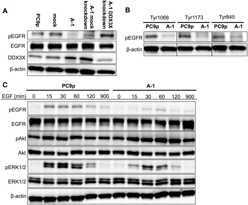
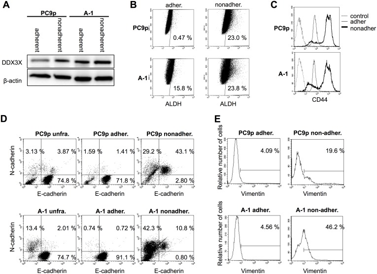
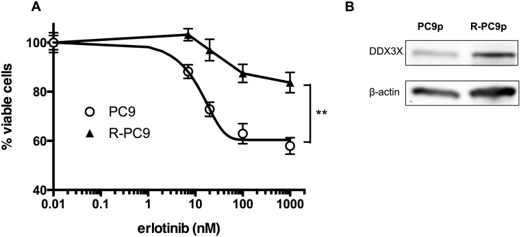
The specific mechanisms how lung cancer cells harboring epidermal growth factor receptor (EGFR) activating mutations can survive treatment with EGFR-tyrosine kinase inhibitors (TKIs) until they eventually acquire treatment-resistance genetic mutations are unclear. The phenotypic diversity of cancer cells caused by genetic or epigenetic alterations (intratumor heterogeneity) confers treatment failure and may foster tumor evolution through Darwinian selection. Recently, we found DDX3X as the protein that was preferentially expressed in murine melanoma with cancer stem cell (CSC)-like phenotypes by proteome analysis. In this study, we transfected PC9, human lung cancer cells harboring EGFR exon19 deletion, with cDNA encoding DDX3X and found that DDX3X, an ATP-dependent RNA helicase, induced CSC-like phenotypes and the epithelial-mesenchymal transition (EMT) accompanied with loss of sensitivity to EGFR-TKI. DDX3X expression was associated with upregulation of Sox2 and increase of cancer cells exhibiting CSC-like phenotypes, such as anchorage-independent proliferation, strong expression of CD44, and aldehyde dehydrogenase (ALDH). The EMT with switching from E-cadherin to N-cadherin was also facilitated by DDX3X. Either ligand-independent or ligand-induced EGFR phosphorylation was inhibited in lung cancer cells that strongly expressed DDX3X. Lack of EGFR signal addiction resulted in resistance to EGFR-TKI. Moreover, we found a small nonadherent subpopulation that strongly expressed DDX3X accompanied by the same stem cell-like properties and the EMT in parental PC9 cells. The unique subpopulation lacked EGFR signaling and was highly resistant to EGFR-TKI. In conclusion, our data indicate that DDX3X may play a critical role for inducing phenotypic diversity, and that treatment targeting DDX3X may overcome primary resistance to EGFR-TKI resulting from intratumor heterogeneity.


Conflict of interest statement

Competing Interests: This study was funded in part by Otsuka Pharmaceutical Co., Ltd. This does not alter the authors’ adherence to PLOS ONE policies on sharing data and materials.

Figures

Figure 1
Figure 1
A. Immunoblotting analysis of DDX3X in parental PC9 cells, mock transfectants, and A-1 cells that were transfected with cDNA encoding DDX3X. B. Immunoblotting analysis of Sox2, KLF4, and c-Myc, in parental PC9 cells, mock transfectants, and A-1 cells. C. Analysis of anchorage-independent cell proliferation by phase-contrast microscopy. D. 1×10⁵ PC9 parental cells or A-1 cells were cultured on 24-well plates for 3 days. Nonadherent cells were collected by gentle rocking in culture medium. The ratio of nonadherent cells was calculated based on the total number of adherent cells. Dead cells were excluded with trypan blue exclusion method. *P<0.05. Data are presented as the mean ± SD of three independent experiments.
Figure 2
Figure 2
A. MTT assay of parental PC9 cells and A-1 cells. Cancer cells were treated with the indicated concentration of erlotinib for 48 h. Samples were prepared in triplicate. Data are presented as the mean ± SD. B. MTT assay of parental PC9 cells, A-1 cells, and A-1 cells with knockdown of DDX3X. Cancer cells were treated with the indicated concentration of erlotinib for 48 h. Samples were prepared in triplicate. Data are presented as the mean ± SD. C. Flow cytometry analysis of cancer cells treated with 20 nM erlotinib for 72 h.
Figure 3
Figure 3
A. A total of 5×10⁴ tumor cells were seeded in 24-well plates on day 0. Twenty-four hours after seeding, erlotinib or vehicle was added at the indicated concentrations. Numbers of live and dead cells were counted with trypan blue staining. % Survival indicates a percentage of live cells, which excluded trypan blue staining, based on total cell number in a well. Samples were prepared in triplicate. Data are presented as the mean ± SD. * is used to indicate significance. B. The %Survival of cancer cells treated with 20 nM erlotinib was demonstrated. A-1, 2, 3, 4, and 5 indicate clone numbers that were transfected with cDNA encoding DDX3X. The %Survival indicates the percentage of live cells that excluded the trypan blue stain, based on total cell number in a well. Samples were prepared in triplicate. Data are presented as the mean ± SD. **p<0.001.
Figure 4
Figure 4
A. Immunoblotting analysis of EGFR, phospho-EGFR (Tyr1068), and DDX3X in cancer cells without EGF supplementation. B. Immunoblotting analysis of phospho-EGFR at Tyr1068, Tyr1173, and Tyr845 in parental PC9 cells and A-1 cells. C. Kinetic analysis of EGFR signaling in parental PC9 cells and A-1 cells. EGFR, phospho-EGFR (Tyr1068), Akt, phospho-Akt, ERK1/2, and phospho-ERK1/2 levels were analyzed by immunoblotting at 0, 15, 30, 60, 120, and 900 min after the addition of 100 ng/mL EGF.
Figure 5
Figure 5
A. Immunoblotting analysis of DDX3X expression in adherent and nonadherent parental PC9 cells and A-1 cells. B. ALDEFLUOR assays were carried out by suspending cells in Aldefluor assay buffer containing 1.5 µM bodipy-aminoacetaldehyde, an ALDH substrate. Cells were then incubated for 45 min at 37°C and analyzed according to the manufacturer’s instructions. C. Flow cytometry analysis of adherent and nonadherent parental PC9 cells and A-1 cells. Histgrams indicate CD44 expression. Dotted lines indicate isotype control, thin lines indicate adherent cells, and the thick lines indicate nonadherent cells. D. Flow cytometry analysis of unfractionated, adherent, and nonadherent cells derived from parental PC9 cells and A-1 cells. X-axis indicates fluorescence intensity of E-cadherin, and Y-axis indicates fluorescence intensity of N-cadherin. E. Flow cytometry analysis of nonadherent cells isolated from parental PC9 and A-1 cells. Histogram plots show vimentin expression on gated N-cadherin+ cells.
Figure 6
Figure 6
A. Immunoblotting analysis of EGFR and phospho-EGFR (Tyr1068) in adherent and nonadherent cancer cells. B. MTT assays were used to measure cell proliferation in adherent and nonadherent PC9 cells and A-1 cells following erlotinib treatment. Data are presented as the mean ± SD. Samples were prepared in triplicate. **P<0.001. C. Flow cytometry analysis with PI staining was used to examine dying cells.
Figure 7
Figure 7
A. MTT assays were used to determine the proliferation of adherent and nonadherent PC9 cells, as well as R-PC9 cells, which were obtained by long-term exposure to increasing concentrations of erlotinib. Data are presented as the mean ± SD. Samples were prepared in triplicate. * indicates significant differences by two-way ANOVA and ad hoc analysis. B. Immunoblotting analysis of DDX3X expression in parental PC9 cells and R-PC9 cells.

Similar articles

Cited by

References

    1. Maemondo M, Inoue A, Kobayashi K, Sugawara S, Oizumi S, et al. (2010) Gefitinib or chemotherapy for non-small-cell lung cancer with mutated EGFR. N Engl J Med 362: 2380–2388. - PubMed
    1. Mitsudomi T, Morita S, Yatabe Y, Negoro S, Okamoto I, et al. (2010) Gefitinib versus cisplatin plus docetaxel in patients with non-small-cell lung cancer harbouring mutations of the epidermal growth factor receptor (WJTOG3405): an open label, randomised phase 3 trial. Lancet Oncol 11: 121–128. - PubMed
    1. Sharma SV, Lee DY, Li B, Quinlan MP, Takahashi F, et al. (2010) A chromatin-mediated reversible drug-tolerant state in cancer cell subpopulations. Cell 141: 69–80. - PMC - PubMed
    1. Lahn BT, Page DC (1997) Functional coherence of the human Y chromosome. Science 278: 675–680. - PubMed
    1. Rocak S, Linder P (2004) DEAD-box proteins: the driving forces behind RNA metabolism. Nat Rev Mol Cell Biol 5: 232–241. - PubMed

Publication types

MeSH terms