

A computer-based automated algorithm for assessing acinar cell loss after experimental pancreatitis
- PMID: 25343460
- PMCID: PMC4208778
- DOI: 10.1371/journal.pone.0110220
A computer-based automated algorithm for assessing acinar cell loss after experimental pancreatitis
Abstract
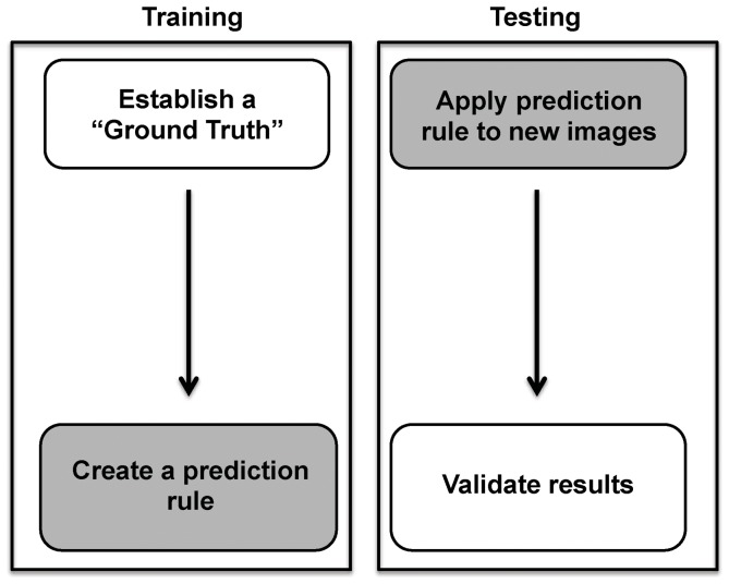
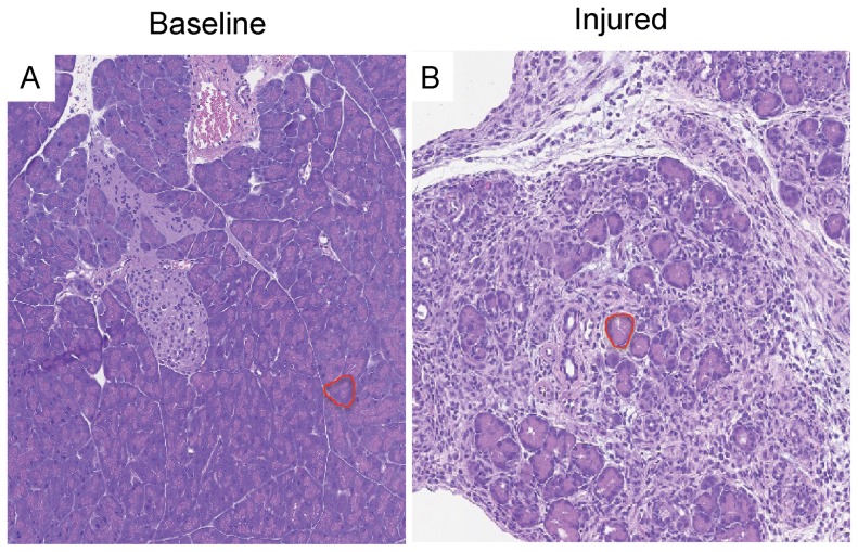
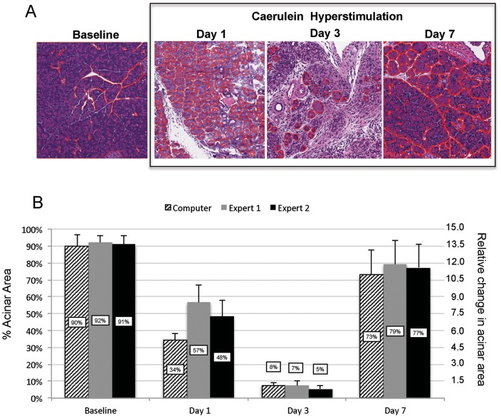
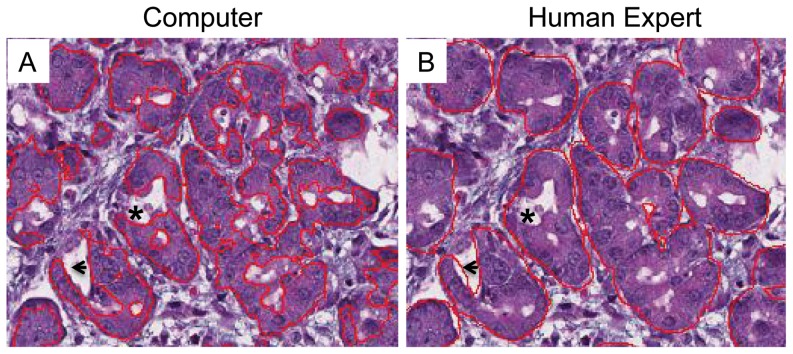
The change in exocrine mass is an important parameter to follow in experimental models of pancreatic injury and regeneration. However, at present, the quantitative assessment of exocrine content by histology is tedious and operator-dependent, requiring manual assessment of acinar area on serial pancreatic sections. In this study, we utilized a novel computer-generated learning algorithm to construct an accurate and rapid method of quantifying acinar content. The algorithm works by learning differences in pixel characteristics from input examples provided by human experts. HE-stained pancreatic sections were obtained in mice recovering from a 2-day, hourly caerulein hyperstimulation model of experimental pancreatitis. For training data, a pathologist carefully outlined discrete regions of acinar and non-acinar tissue in 21 sections at various stages of pancreatic injury and recovery (termed the "ground truth"). After the expert defined the ground truth, the computer was able to develop a prediction rule that was then applied to a unique set of high-resolution images in order to validate the process. For baseline, non-injured pancreatic sections, the software demonstrated close agreement with the ground truth in identifying baseline acinar tissue area with only a difference of 1% ± 0.05% (p = 0.21). Within regions of injured tissue, the software reported a difference of 2.5% ± 0.04% in acinar area compared with the pathologist (p = 0.47). Surprisingly, on detailed morphological examination, the discrepancy was primarily because the software outlined acini and excluded inter-acinar and luminal white space with greater precision. The findings suggest that the software will be of great potential benefit to both clinicians and researchers in quantifying pancreatic acinar cell flux in the injured and recovering pancreas.
Conflict of interest statement
Figures

Similar articles
-
Ca2+ Influx Channel Inhibitor SARAF Protects Mice From Acute Pancreatitis.Gastroenterology. 2019 Dec;157(6):1660-1672.e2. doi: 10.1053/j.gastro.2019.08.042. Epub 2019 Sep 4. Gastroenterology. 2019. PMID: 31493399
-
Local hyperthyroidism promotes pancreatic acinar cell proliferation during acute pancreatitis.J Pathol. 2019 Jun;248(2):217-229. doi: 10.1002/path.5247. Epub 2019 Apr 4. J Pathol. 2019. PMID: 30714146
-
Mnk1 is a novel acinar cell-specific kinase required for exocrine pancreatic secretion and response to pancreatitis in mice.Gut. 2015 Jun;64(6):937-47. doi: 10.1136/gutjnl-2013-306068. Epub 2014 Jul 18. Gut. 2015. PMID: 25037190
-
The MET Receptor Tyrosine Kinase Confers Repair of Murine Pancreatic Acinar Cells following Acute and Chronic Injury.PLoS One. 2016 Oct 31;11(10):e0165485. doi: 10.1371/journal.pone.0165485. eCollection 2016. PLoS One. 2016. PMID: 27798657 Free PMC article.
-
Regeneration and repair of the exocrine pancreas.Annu Rev Physiol. 2015;77:229-49. doi: 10.1146/annurev-physiol-021014-071727. Epub 2014 Oct 24. Annu Rev Physiol. 2015. PMID: 25386992 Free PMC article. Review.
Cited by
-
Loss of Sirt2 increases and prolongs a caerulein-induced pancreatitis permissive phenotype and induces spontaneous oncogenic Kras mutations in mice.Sci Rep. 2018 Nov 7;8(1):16501. doi: 10.1038/s41598-018-34792-y. Sci Rep. 2018. PMID: 30405152 Free PMC article.
-
Valproic Acid Limits Pancreatic Recovery after Pancreatitis by Inhibiting Histone Deacetylases and Preventing Acinar Redifferentiation Programs.Am J Pathol. 2015 Dec;185(12):3304-15. doi: 10.1016/j.ajpath.2015.08.006. Epub 2015 Oct 23. Am J Pathol. 2015. PMID: 26476347 Free PMC article.
-
A supervised learning framework for pancreatic islet segmentation with multi-scale color-texture features and rolling guidance filters.Cytometry A. 2016 Oct;89(10):893-902. doi: 10.1002/cyto.a.22929. Epub 2016 Aug 25. Cytometry A. 2016. PMID: 27560544 Free PMC article.
-
Pancreatic gene expression during recovery after pancreatitis reveals unique transcriptome profiles.Sci Rep. 2018 Jan 23;8(1):1406. doi: 10.1038/s41598-018-19392-0. Sci Rep. 2018. PMID: 29362419 Free PMC article.
References
-
- Bhatia M (2004) Apoptosis versus necrosis in acute pancreatitis. Am J Physiol Gastrointest Liver Physiol 286: G189–196. - PubMed
-
- Bhatia M, Wong FL, Cao Y, Lau HY, Huang J, et al. (2005) Pathophysiology of acute pancreatitis. Pancreatology 5: 132–144. - PubMed
-
- Gukovskaya AS, Gukovsky I, Jung Y, Mouria M, Pandol SJ (2002) Cholecystokinin induces caspase activation and mitochondrial dysfunction in pancreatic acinar cells. Roles in cell injury processes of pancreatitis. J Biol Chem 277: 22595–22604. - PubMed
-
- Jensen JN, Cameron E, Garay MV, Starkey TW, Gianani R, et al. (2005) Recapitulation of elements of embryonic development in adult mouse pancreatic regeneration. Gastroenterology 128: 728–741. - PubMed
Publication types
MeSH terms
Substances
Grants and funding
LinkOut - more resources
Full Text Sources
Other Literature Sources
Medical
