. 2014 Oct 24;9(10):e109674.
doi: 10.1371/journal.pone.0109674. eCollection 2014.

Molecular insights into the interaction between Plasmodium falciparum apical membrane antigen 1 and an invasion-inhibitory peptide

Affiliations

Molecular insights into the interaction between Plasmodium falciparum apical membrane antigen 1 and an invasion-inhibitory peptide

Geqing Wang et al. PLoS One. .

Abstract

Apical membrane antigen 1 (AMA1) of the human malaria parasite Plasmodium falciparum has been implicated in invasion of the host erythrocyte. It interacts with malarial rhoptry neck (RON) proteins in the moving junction that forms between the host cell and the invading parasite. Agents that block this interaction inhibit invasion and may serve as promising leads for anti-malarial drug development. The invasion-inhibitory peptide R1 binds to a hydrophobic cleft on AMA1, which is an attractive target site for small molecules that block parasite invasion. In this work, truncation and mutational analyses show that Phe5-Phe9, Phe12 and Arg15 in R1 are the most important residues for high affinity binding to AMA1. These residues interact with two well-defined binding hot spots on AMA1. Computational solvent mapping reveals that one of these hot spots is suitable for small molecule targeting. We also confirm that R1 in solution binds to AMA1 with 1:1 stoichiometry and adopts a secondary structure consistent with the major form of R1 observed in the crystal structure of the complex. Our results provide a basis for designing high affinity inhibitors of the AMA1-RON2 interaction.


Conflict of interest statement

Competing Interests: The authors have declared that no competing interests exist.

Figures

Figure 1
Figure 1. Identification of the minimal binding construct of R1 peptide.
A. Amino acid sequences of PfRON22031–2042, native R1 and truncated peptides. Residues that are conserved between R1 and RON2 are highlighted in red. B. Co-crystal structure of PfAMA1 bound to R1 peptide (PDB ID: 3SRJ, [27]). AMA1 is presented as a grey surface; R1 is presented as a cartoon (the minimal binding construct Phe5-Met16 is shown in blue, Val1-Glu4 and His17-Ile18 are in yellow). Side chains of the conserved residues are highlighted in red. The minor form of R1 is omitted in this structure. C. Structural comparison of R15–16 (blue) and PfRON22031–2042 (orange) bound to AMA1. The structure of the R1 peptide bound to PfAMA1 (PDB ID: 3SRJ) superimposed onto the co-crystal structure of PfAMA1-PfRON2 (PDB ID: 3ZWZ, [27]). Only Phe5-Met16 of R1 and Ala2031-Met2042 of PfRON2 are shown for clarity.
Figure 2
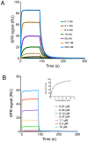
Figure 2. SPR analysis of peptides binding to immobilized 3D7 PfAMA1104–442.
A series of concentrations, as indicated in sensorgrams, of native R1 (panel A) and truncated R15–16 (panel B) was injected over the AMA1-immobilized surface. k a and k d of native R1 were determined by globally fitting to a 1∶1 binding model. The apparent equilibrium dissociation constants K D for other peptides were determined using a steady-state affinity model and are given in Table 1.
Figure 3
Figure 3. Comparison of the 1H-15N-HSQC spectra of f-2H, u-13C, 15N-labelled R1 in the absence (blue) and presence (red) of a saturating concentration of fractionally deuterated 3D7 PfAMA1104–442 at pH 7 and 40°C.
Some amide resonances (Ala3, Phe9, Ser10, Lys11, Phe12, Gly13, Ser14 and Arg15) of free R1 were broadened beyond detection at pH 7 and 40°C and their predicted resonances are indicated as black circles in the spectrum (prediction was made as described in the text). N/H indicates unassigned amide resonances of bound R1.
Figure 4
Figure 4. Elution profile of AMA1 in the absence (blue) and presence (red) of R1 peptide on an analytical size exclusion column.
The elution time of bovine serum albumin (BSA) was determined for the size exclusion column that was used to elute AMA1 (Superdex 75 HR 10/30, column dimension 1.0×30 cm, column volume 23.6 mL). Both apo AMA1 (200 µM) and R1-bound AMA1 (200 µM AMA1+250 µM R1) showed a similar elution profile. The peak eluting at 10.5 mL is consistent with monomeric AMA1 (MW 41.3 kDa when His tag is not cleaved). SDS-PAGE (inset) confirms only one protein band corresponding to AMA1 is present. NR = non-reducing, R = reducing. The peak at ∼14 mL results from the addition of Complete protease inhibitor cocktail (Roche) to the buffer (Figure S8 in File S1), and was also verified by mass spectroscopy.
Figure 5
Figure 5. SAXS analysis of AMA1 alone and AMA1 in the presence of R1.
(A) AMA1 scattering data fitted to the crystal structure of AMA1 (PDB 1Z40) (Chi-square = 0.61). (B) AMA1+R1 scattering data fitted to the crystal structure of AMA1 bound to R1 (PDB 3SRJ) (Chi-square = 0.72). Q is the momentum transfer vector.
Figure 6
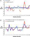
Figure 6. NMR Cα secondary chemical shifts.
A. The Cα secondary shifts for free (blue) and AMA1-bound (red) R1 at pH 7 and 40°C. B. Comparison of the experimentally determined Cα secondary shifts of bound R1 (red) and Cα secondary shifts predicted for the major form of R1 bound to AMA1 in the crystal structure (Chain C, PDB ID: 3SRJ) using SHIFTX2 (blue) .
Figure 7
Figure 7. Computational solvent mapping of AMA1 using FTMAP.
(A) Mapping results for R1-bound 3D7 PfAMA1 (grey, PDB ID: 3SRJ). R1 peptides and water were removed prior to mapping. The five largest consensus sites, CS1 (cyan, 21 probe clusters), CS2 (magenta, 17 probe clusters), CS3 (yellow, 13 probe clusters), CS4 (salmon, 10 probe clusters) and CS5 (grey, 9 probe clusters) are located in a large hydrophobic pocket that binds to the Phe5-Phe9 segment of R1 peptide. The position of the R1 peptide in the crystal structure is shown for reference (blue). Phe5-Phe9 side chains are displayed as sticks and labelled individually as shown in the figure. The surface of the pocket is coloured according to side chain colours in panel B. (B) The residues that interact with small molecule probe clusters in the pocket are shown as sticks (green/orange). A cluster of five Tyr residues is highlighted in orange. Broken purple line indicates the displaced domain II loop in the R1-bound conformation. (C) Mapping results for IgNAR-bound 3D7 PfAMA1 (grey, PDB ID: 2Z8V). IgNAR and water were removed prior to mapping. Two consensus sites CS3 (yellow, 14 probe clusters) and CS5 (grey, 9 probe clusters) are located in a domain II loop-protected pocket. The surface of the protein is coloured according to colour scheme in panel D. An inset highlights probes that fit into a deep, narrow pocket. (D) Some key interacting residues (highlighted as green/orange sticks), which bind probes when the domain II loop is displaced (panel B), are still solvent accessible to small molecule organic probes when the pocket is partially protected by the domain II loop (purple).

References

    1. World Health Organisation (2013) World Malaria Report 2013.
    1. MacRaild CA, Anders RF, Foley M, Norton RS (2011) Apical membrane antigen 1 as an anti-malarial drug target. Curr Top Med Chem 11: 2039–2047. - PubMed
    1. Miller LH, Baruch DI, Marsh K, Doumbo OK (2002) The pathogenic basis of malaria. Nature 415: 673–679. - PubMed
    1. Srinivasan P, Beatty WL, Diouf A, Herrera R, Ambroggio X, et al. (2011) Binding of Plasmodium merozoite proteins RON2 and AMA1 triggers commitment to invasion. Proc Natl Acad Sci U S A 108: 13275–13280. - PMC - PubMed
    1. Tonkin ML, Roques M, Lamarque MH, Pugniere M, Douguet D, et al. (2011) Host cell invasion by apicomplexan parasites: insights from the co-structure of AMA1 with a RON2 peptide. Science 333: 463–467. - PubMed

Publication types

MeSH terms

LinkOut - more resources