. 2015 Mar;21(5-6):1024-35.
doi: 10.1089/ten.TEA.2014.0022. Epub 2015 Feb 5.

Transplantation of heterospheroids of islet cells and mesenchymal stem cells for effective angiogenesis and antiapoptosis

Affiliations

Transplantation of heterospheroids of islet cells and mesenchymal stem cells for effective angiogenesis and antiapoptosis

Jung-Youn Shin et al. Tissue Eng Part A. 2015 Mar.

Abstract

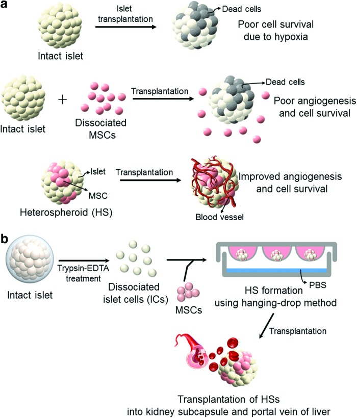
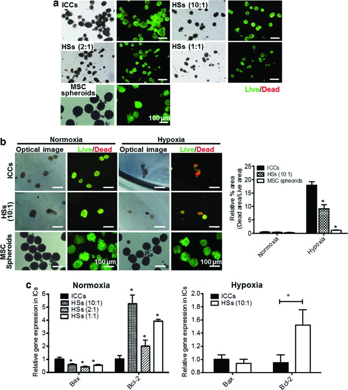
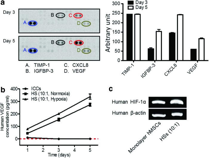
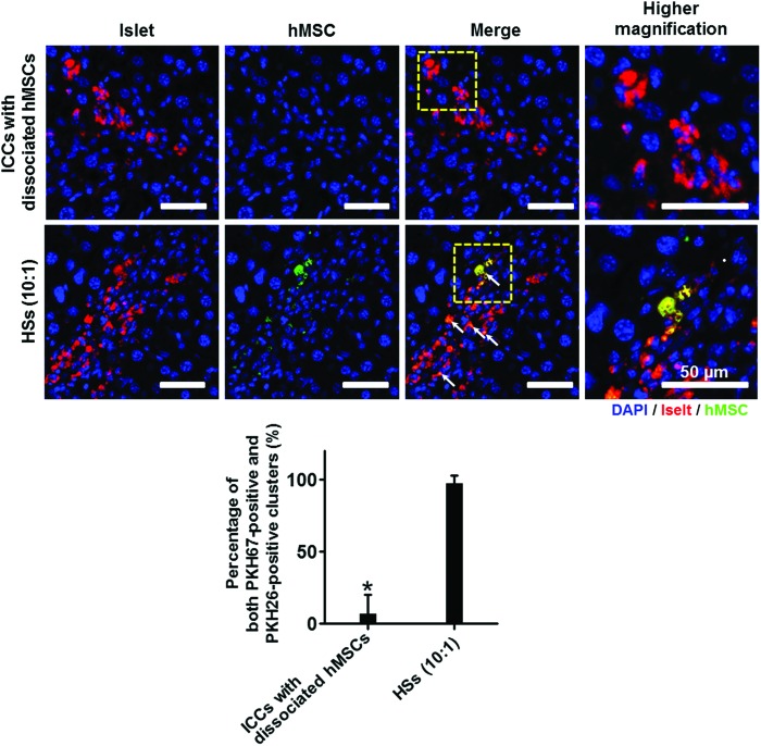
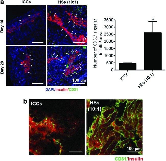
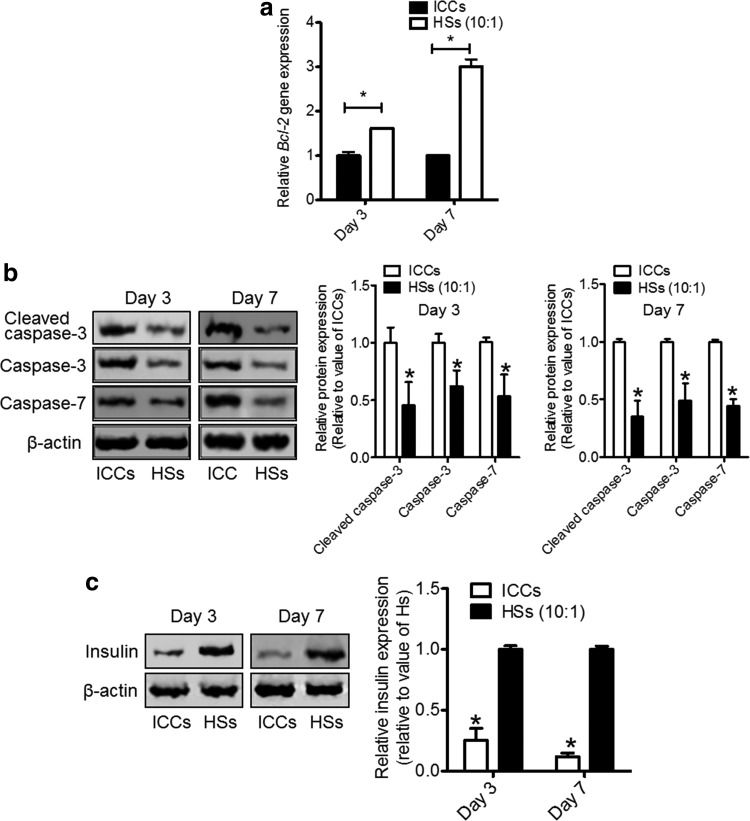
Although islet transplantation has been suggested as an alternative therapy for type 1 diabetes, there are efficiency concerns that are attributed to poor engraftment of transplanted islets. Hypoxic condition and delayed vasculogenesis induce necrosis and apoptosis of the transplanted islets. To overcome these limitations in islet transplantation, heterospheroids (HSs), which consist of rat islet cells (ICs) and human bone marrow-derived mesenchymal stem cells (hMSCs), were transplanted to the kidney and liver. The HSs cultured under the hypoxic condition system exhibited a significant increase in antiapoptotic gene expression in ICs. hMSCs in the HSs secreted angiogenic and antiapoptotic proteins. With the HS system, ICs and hMSCs were successfully located in the same area of the liver after transplantation of HSs through the portal vein, whereas the transplantation of islets and the dissociated hMSCs did not result in localization of transplanted ICs and hMSCs in the same area. HS transplantation resulted in an increase in angiogenesis at the transplantation area and a decrease in the apoptosis of transplanted ICs after transplantation into the kidney subcapsule compared with transplantation of islet cell clusters (ICCs). Insulin production levels of ICs were higher in the HS transplantation group compared with the ICC transplantation group. The HS system may be a more efficient transplantation method than the conventional methods for the treatment of type 1 diabetes.


Figures

<b>FIG. 1.</b>
FIG. 1.
Schematic illustration showing (a) an efficient method of cotransplantation of islets and MSCs for improved angiogenesis and IC survival and (b) a protocol for the formation of HSs of ICs and MSCs. (a) Islet transplantation often results in poor islet survival due to hypoxia in the transplantation region. Cotransplantation with MSCs can induce angiogenesis and improve IC survival. Importantly, transplantation of HSs of ICs and MSCs would be more effective for locating ICs with MSCs, angiogenesis, and IC survival than in the transplantation of islets and dissociated MSCs. (b) HS consisting of rat ICs and hMSCs can be prepared using the hanging-drop method. hMSCs, human bone marrow-derived mesenchymal stem cells; HSs, heterospheroids; IC, islet cell; MSCs, mesenchymal stem cells. Color images available online at www.liebertpub.com/tea
<b>FIG. 2.</b>
FIG. 2.
Characteristic analysis of ICCs and HSs after complete aggregation. (a) Light microscopic (left) and live/dead cell (right) images of ICCs and HSs. Green and red signals indicate live and dead cells, respectively. Scale bars=100 μm. (b) The diameters of ICCs and HSs with various ICs/hMSCs ratios of 10:1, 2:1, and 1:1. There was no statistical significance between any two groups. (c) Confocal laser scanning microscopic images of cell clusters (green: ICs; red: hMSCs). Scale bars=50 μm. (d) Cell images after dissociation of HSs into ICs and hMSCs (left panel, blue: DAPI, red: hMSCs) and cell composition (right panel). Scale bars=100 μm. DAPI, 4′,6-diamidino-2-phenylindole; ICCs, islet cell clusters. Color images available online at www.liebertpub.com/tea
<b>FIG. 3.</b>
FIG. 3.
In vitro viability and apoptosis of cultured HSs, ICCs, and hMSC spheroids. (a) Long-term in vitro viability of ICCs, HSs, and hMSC spheroids on day 7 as evaluated by live/dead assay. Scale bars=100 μm. (b) Live/dead images (left panel) and their relative quantification graph (right panel) of ICCs, HSs (10:1), and hMSC spheroids cultured under normoxic and hypoxic conditions for 2 days. Green and red signals indicate live and dead cells, respectively. Scale bars=100 μm. *p<0.05 compared with ICCs. (c) Relative expression of Bax and Bcl-2 of ICs in ICCs or HSs cultured under normoxic or hypoxic condition, as evaluated by qRT-PCR. *p<0.05 compared with ICCs. qRT-PCR, quantitative reverse transcriptase-polymerase chain reaction. Color images available online at www.liebertpub.com/tea
<b>FIG. 4.</b>
FIG. 4.
Analysis of angiogenic factors present in cell culture supernate, and human HIF-1α mRNA expressions in monolayer hMSCs and hMSCs of HSs on day 3. (a) The relative amounts of human angiogenic factors (TIMP-1, IGFBP-3, CXCL8, and VEGF) present in cell culture supernate of HSs (10:1). Cell culture supernate was collected on days 3 and 5 and subjected to a human angiogenesis antibody array. Since the data represent the average values from a single test, which yielded two test values for each individual cytokine, statistical significance was not determined. (b) The amounts of hVEGF secreted from rat ICCs and HSs (10:1) containing hMSCs and rat ICs cultured in normoxic conditions and HSs (10:1) cultured in hypoxic conditions at various culture time points, as evaluated by ELISA. The red dashed line indicates 0 pg/ml of hVEGF. p<0.05 between any two groups at all time points. (c) Human HIF-1α mRNA expressions of monolayer hMSCs or HSs on day 3. CXCL8, interleukin-8; ELISA, enzyme-linked immunosorbent assay; HIF-1α, hypoxia inducible factor-1α; hVEGF, human vascular endothelial growth factor; IGFBP-3, insulin-like growth factor-binding protein-3; TIMP-1, tissue inhibitor of metalloproteinase-1; VEGF, vascular endothelial growth factor. Color images available online at www.liebertpub.com/tea
<b>FIG. 5.</b>
FIG. 5.
ICs and hMSC localization in the portal vein of the liver. Before transplantation, ICs and hMSCs were labeled with PKH26 (red) and PKH67 (green), respectively. The livers were harvested 7 days after transplantation. Arrows indicate the ICs localized with hMSCs in the liver. Blue indicates nuclei stained with DAPI. Scale bars=50 μm. The percentages of both PKH67-positive (hMSC) and PKH26-positive (IC) clusters were evaluated from total clusters found in each liver (lower panel). *p<0.05 compared with HSs (10:1) group. Color images available online at www.liebertpub.com/tea
<b>FIG. 6.</b>
FIG. 6.
Angiogenesis at the transplantation area in the kidney subcapsule after transplantation of both ICCs and HSs (IC:hMSC=10:1). (a) CD31-positive (green) microvessels with insulin-positive (red) transplanted ICs (left panel). The kidneys that contained transplanted islets were harvested 14 and 28 days after the transplantation. Arrows indicate CD31-positive microvessels in the transplantation region. Blue indicates nuclei stained with DAPI. Scale bars=100 μm. Right panel. The density of CD31-positive microvessels in the insulin-positive region. *p<0.05 compared with ICCs. (b) Whole-mount immunostaining of CD31 (green) and insulin (red) in the transplantation area. The kidneys containing transplanted islets were harvested 28 days after the transplantation. Scale bars=100 μm. Color images available online at www.liebertpub.com/tea
<b>FIG. 7.</b>
FIG. 7.
Evaluation of apoptosis and insulin content of ICCs and ICs in HSs in the transplantation area of the kidneys. (a) Relative expression of rat-specific Bcl-2 of ICCs and ICs in HSs (10:1) as evaluated by qRT-PCR. *p<0.05 compared with ICCs. (b) Apoptotic protein expression was evaluated by western blot (upper panel) using antibodies against cleaved caspase-3, caspase-3, and caspase-7, and their protein expression levels were evaluated (lower panel). *p<0.05 compared with ICCs. (c) Insulin content was evaluated by western blot (left panel) using antibodies against insulin, and their protein expression levels were evaluated (right panel). *p<0.05 compared with HSs.

References

    1. Shapiro A.M., Ricordi C., Hering B.J., Auchincloss H., Lindblad R., Robertson R.P., et al. . International trial of the Edmonton protocol for islet transplantation. N Engl J Med 355,1318, 2006 - PubMed
    1. Jeong J.H., Hong S.W., Hong S., Yook S., Jung Y., Park J.B., et al. . Surface camouflage of pancreatic islets using 6-arm-PEG-catechol in combined therapy with tacrolimus and anti-CD154 monoclonal antibody for xenotransplantation. Biomaterials 32,7961, 2011 - PubMed
    1. Gibly R.F., Graham J.G., Luo X., Lowe W.L., Jr., Hering B.J., and Shea L.D.Advancing islet transplantation: from engraftment to the immune response. Diabetologia 54,2494, 2011 - PMC - PubMed
    1. Goto M., Tjernberg J., Dufrane D., Elgue G., Brandhorst D., Ekdahl K.N., et al. . Dissecting the instant blood-mediated inflammatory reaction in islet xenotransplantation. Xenotransplantation 15,225, 2008 - PMC - PubMed
    1. Bennet W., Groth C.G., Larsson R., Nilsson B., and Korsgren O.Isolated human islets trigger an instant blood mediated inflammatory reaction: implications for intraportal islet transplantation as a treatment for patients with type 1 diabetes. Ups J Med Sci 105,125, 2000 - PubMed

Publication types

MeSH terms