

Characterization and evaluation of the artemis camera for fluorescence-guided cancer surgery
- PMID: 25344146
- PMCID: PMC4422838
- DOI: 10.1007/s11307-014-0799-z
Characterization and evaluation of the artemis camera for fluorescence-guided cancer surgery
Abstract
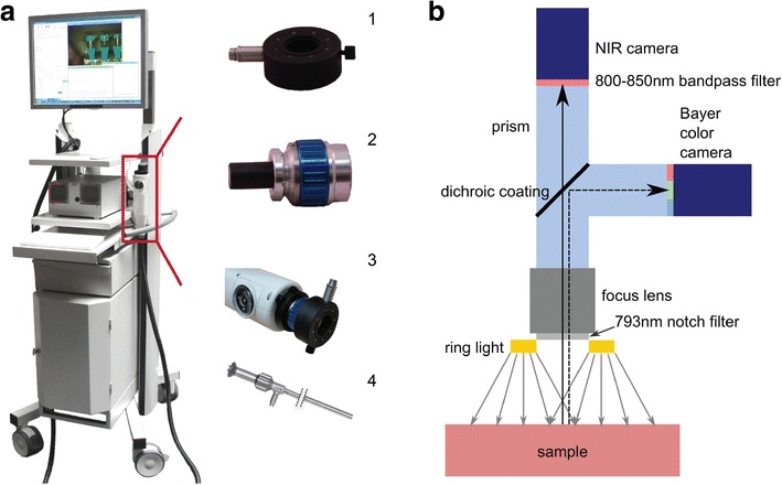
Purpose: Near-infrared (NIR) fluorescence imaging can provide the surgeon with real-time visualization of, e.g., tumor margins and lymph nodes. We describe and evaluate the Artemis, a novel, handheld NIR fluorescence camera.
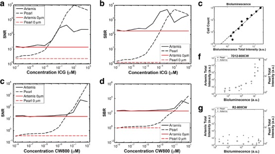
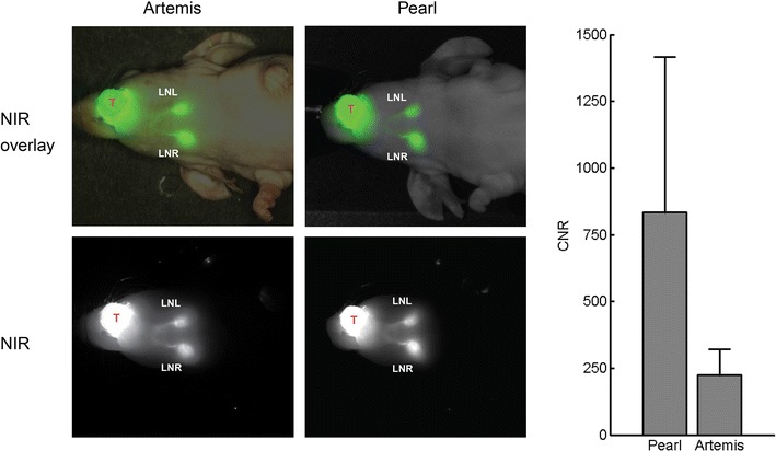
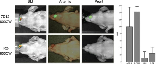
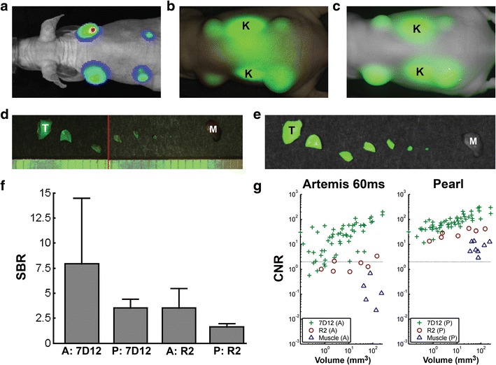
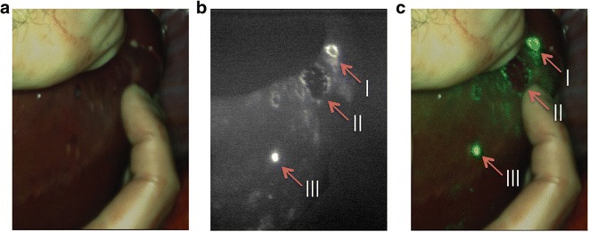
Procedures: We evaluated minimal detectable cell numbers (FaDu-luc2, 7D12-IRDye 800CW), preclinical intraoperative detection of sentinel lymph nodes (SLN) using indocyanine green (ICG), and of orthotopic tongue tumors using 7D12-800CW. Results were compared with the Pearl imager. Clinically, three patients with liver metastases were imaged using ICG.
Results: Minimum detectable cell counts for Artemis and Pearl were 2 × 10(5) and 4 × 10(4) cells, respectively. In vivo, seven SLNs were detected in four mice with both cameras. Orthotopic OSC-19-luc2-cGFP tongue tumors were clearly identifiable, and a minimum FaDu-luc2 tumor size of 1 mm(3) could be identified. Six human malignant lesions were identified during three liver surgery procedures.
Conclusions: Based on this study, the Artemis system has demonstrated its utility in fluorescence-guided cancer surgery.
Conflict of interest statement
The authors report no conflicts of interest
Figures
Similar articles
-
Intraoperative fluorescence delineation of head and neck cancer with a fluorescent anti-epidermal growth factor receptor nanobody.Int J Cancer. 2014 Jun 1;134(11):2663-73. doi: 10.1002/ijc.28601. Epub 2013 Dec 12. Int J Cancer. 2014. PMID: 24222574 Free PMC article.
-
Identification of metastatic nodal disease in a phase 1 dose-escalation trial of intraoperative sentinel lymph node mapping in non-small cell lung cancer using near-infrared imaging.J Thorac Cardiovasc Surg. 2013 Sep;146(3):562-70; discussion 569-70. doi: 10.1016/j.jtcvs.2013.04.010. Epub 2013 Jun 19. J Thorac Cardiovasc Surg. 2013. PMID: 23790404 Free PMC article. Clinical Trial.
-
Near-infrared fluorescence sentinel lymph node mapping of the oral cavity in head and neck cancer patients.Oral Oncol. 2013 Jan;49(1):15-9. doi: 10.1016/j.oraloncology.2012.07.017. Epub 2012 Aug 28. Oral Oncol. 2013. PMID: 22939692 Free PMC article.
-
Sentinel node mapping with indocyanine green and endoscopic near-infrared fluorescence imaging in endometrial cancer. A pilot study and review of the literature.Gynecol Oncol. 2015 Jun;137(3):443-7. doi: 10.1016/j.ygyno.2015.03.004. Epub 2015 Mar 11. Gynecol Oncol. 2015. PMID: 25771495 Review.
-
Diagnostic value of indocyanine green fluorescence guided sentinel lymph node biopsy in vulvar cancer: A systematic review.Gynecol Oncol. 2021 May;161(2):436-441. doi: 10.1016/j.ygyno.2021.01.031. Epub 2021 Feb 5. Gynecol Oncol. 2021. PMID: 33551201
Cited by
-
Intraoperative imaging of folate receptor alpha positive ovarian and breast cancer using the tumor specific agent EC17.Oncotarget. 2016 May 31;7(22):32144-55. doi: 10.18632/oncotarget.8282. Oncotarget. 2016. PMID: 27014973 Free PMC article. Clinical Trial.
-
Validation of a Three-Dimensional Head and Neck Spheroid Model to Evaluate Cameras for NIR Fluorescence-Guided Cancer Surgery.Int J Mol Sci. 2021 Feb 17;22(4):1966. doi: 10.3390/ijms22041966. Int J Mol Sci. 2021. PMID: 33671198 Free PMC article.
-
NIR fluorescence-guided tumor surgery: new strategies for the use of indocyanine green.Int J Nanomedicine. 2019 Sep 25;14:7823-7838. doi: 10.2147/IJN.S207486. eCollection 2019. Int J Nanomedicine. 2019. PMID: 31576126 Free PMC article. Review.
-
Margin Analysis in Head and Neck Cancer: State of the Art and Future Directions.Ann Surg Oncol. 2019 Nov;26(12):4070-4080. doi: 10.1245/s10434-019-07645-9. Epub 2019 Aug 5. Ann Surg Oncol. 2019. PMID: 31385128 Free PMC article. Review.
-
EpCAM as multi-tumour target for near-infrared fluorescence guided surgery.BMC Cancer. 2016 Nov 14;16(1):884. doi: 10.1186/s12885-016-2932-7. BMC Cancer. 2016. PMID: 27842504 Free PMC article.
References
Publication types
MeSH terms
Substances
LinkOut - more resources
Full Text Sources
Other Literature Sources
Miscellaneous
