

Anal sphincter complex: 2D and 3D endoanal and translabial ultrasound measurement variation in normal postpartum measurements
- PMID: 25344221
- PMCID: PMC4578150
- DOI: 10.1007/s00192-014-2524-5
Anal sphincter complex: 2D and 3D endoanal and translabial ultrasound measurement variation in normal postpartum measurements
Abstract
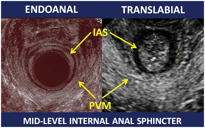
Introduction and hypothesis: Women may experience anal sphincter anatomy changes after vaginal birth (VB) or Cesarean delivery (CD). Therefore, accurate and acceptable imaging options to evaluate the anal sphincter complex (ASC) are needed. ASC measurements may differ between translabial (TLUS) and endoanal (EAUS) ultrasound imaging and between 2D and 3D US. The objective of this analysis was to describe measurement variation between these modalities.
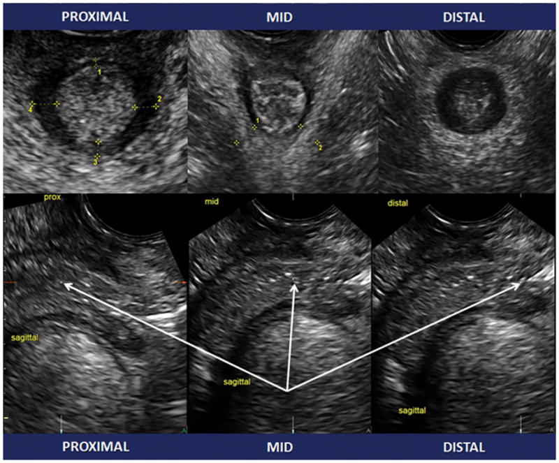
Methods: Primiparous women underwent 2D and 3D TLUS imaging of the ASC 6 months after VB or CD. A subset of women also underwent EAUS measurements. Measurements included internal anal sphincter (IAS) thickness at proximal, mid, and distal levels and the external anal sphincter (EAS) at 3, 6, 9, and 12 o'clock positions, as well as bilateral thickness of the pubovisceralis muscle (PVM).
Results: There were 433 women presenting for US: 423 had TLUS and 64 had both TLUS and EAUS of the ASC. All IAS measurements were significantly thicker on TLUS than EAUS (all p < 0.01), while EAS measurements were significantly thicker on EAUS (p < 0.01). PVM measurements with 3D or 2D imaging were similar (p > 0.20). On both TLUS and EAUS, there were multiple sites where significant asymmetry existed in left versus right measurements.
Conclusions: US modality used to image the ASC introduces small but significant changes in measurements, and the direction of the bias depends on the muscle and location being imaged.
Conflict of interest statement
KV Meriwether: No conflicts of interest to disclose
RJ Hall: No conflicts of interest to disclose
LM Leeman: No conflicts of interest to disclose
L Migliaccio: No conflicts of interest to disclose
C Qualls: No conflicts of interest to disclose
RG Rogers: Chair DSMB for the Transform trial sponsored by American Medical Systems
Figures

References
-
- DeLancey JO. The hidden epidemic of pelvic floor dysfunction: achievable goals for improved prevention and treatment. Am J Obstet Gynecol. 2005 May;192(5):1488–95. - PubMed
-
- Oberwalder M, Connor J, Wexner SD. Meta-analysis to determine the incidence of obstetric anal sphincter damage. Br J Surg. 2003 Nov;90(11):1333–7. - PubMed
-
- Faltin DL, Boulvain M, Floris LA, Irion O. Diagnosis of anal sphincter tears to prevent fecal incontinence: a randomized controlled trial. Obstet Gynecol. 2005 Jul;106(1):6–13. - PubMed
MeSH terms
Grants and funding
LinkOut - more resources
Full Text Sources
Other Literature Sources
Medical
Miscellaneous
