. 2015 Jan;81(1):332-8.
doi: 10.1128/AEM.02573-14. Epub 2014 Oct 24.

Salivary mucins protect surfaces from colonization by cariogenic bacteria

Affiliations

Salivary mucins protect surfaces from colonization by cariogenic bacteria

Erica Shapiro Frenkel et al. Appl Environ Microbiol. 2015 Jan.

Abstract

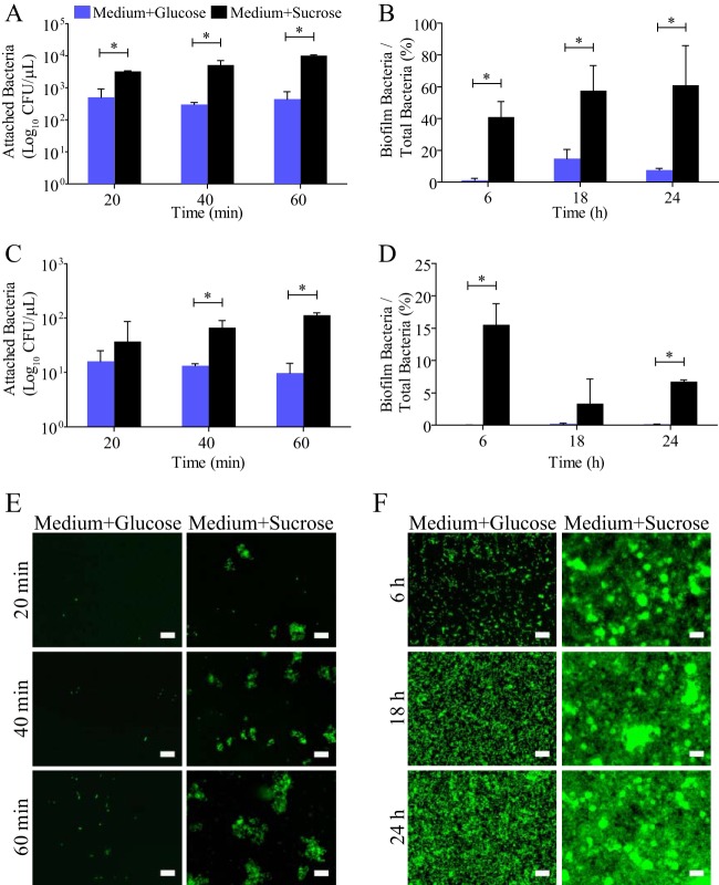
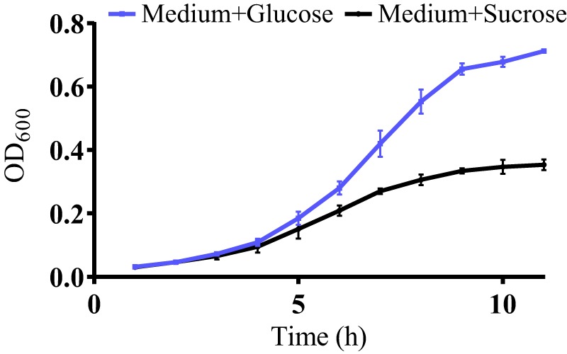
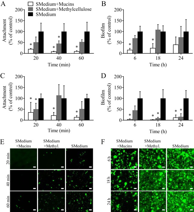
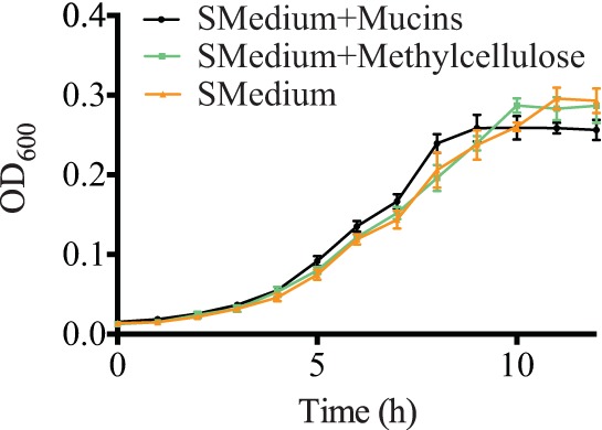
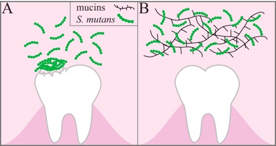
Understanding how the body's natural defenses function to protect the oral cavity from the myriad of bacteria that colonize its surfaces is an ongoing topic of research that can lead to breakthroughs in treatment and prevention. One key defense mechanism on all moist epithelial linings, such as the mouth, gastrointestinal tract, and lungs, is a layer of thick, well-hydrated mucus. The main gel-forming components of mucus are mucins, large glycoproteins that play a key role in host defense. This study focuses on elucidating the connection between MUC5B salivary mucins and dental caries, one of the most common oral diseases. Dental caries is predominantly caused by Streptococcus mutans attachment and biofilm formation on the tooth surface. Once S. mutans attaches to the tooth, it produces organic acids as metabolic by-products that dissolve tooth enamel, leading to cavity formation. We utilize CFU counts and fluorescence microscopy to quantitatively show that S. mutans attachment and biofilm formation are most robust in the presence of sucrose and that aqueous solutions of purified human MUC5B protect surfaces by acting as an antibiofouling agent in the presence of sucrose. In addition, we find that MUC5B does not alter S. mutans growth and decreases surface attachment and biofilm formation by maintaining S. mutans in the planktonic form. These insights point to the importance of salivary mucins in oral health and lead to a better understanding of how MUC5B could play a role in cavity prevention or diagnosis.


Figures

FIG 1
FIG 1
Sucrose enhances S. mutans attachment and biofilm formation. The levels of S. mutans attachment (A) and biofilm formation (B) on glass are significantly enhanced at all time points when the bacteria are grown in BHI containing 1% sucrose (Medium+Sucrose) compared to the levels achieved in BHI containing 1% glucose (Medium+Glucose). S. mutans attachment (C) and biofilm formation (D) on hydroxyapatite are similarly increased in the presence of sucrose, illustrating that the effect of sucrose on S. mutans physiology is not surface specific. Fluorescence microscopy images verify the findings of the CFU count experiments by showing an increase in S. mutans attachment (E) and biofilm formation (F) on glass in the presence of sucrose. *, statistically significant difference determined by Student's t test (P < 0.02). Error bars represent SDs. Scale bars, 20 μm.
FIG 2
FIG 2
Supplemental sugar alters S. mutans growth. A growth curve of S. mutans in BHI with added 1% glucose (Medium+Glucose) or 1% sucrose (Medium+Sucrose) shows that S. mutans' growth rate changes based on the specific sugar present in the growth medium. Error bars represent SDs.
FIG 3
FIG 3
Salivary mucins reduce S. mutans attachment and biofilm formation. The addition of 0.3% mucins to the control medium, BHI containing 1% sucrose (SMedium), significantly reduces the levels of S. mutans attachment and biofilm formation on glass (A, B) and hydroxyapatite (C, D) compared to the levels obtained with the control consisting of BHI with 1% sucrose. Similarly, the addition of 0.3% methylcellulose to BHI with 1% sucrose reduces S. mutans attachment and biofilm formation; however, the effect is not significant for the majority of time points studied. Fluorescence microscopy was used to visually assess S. mutans attachment (E) and biofilm formation (F) on glass when the bacteria are grown in BHI with 1% sucrose and 0.3% mucins, BHI with 1% sucrose and 0.3% methylcellulose (Methyl.), and BHI with 1% sucrose. *, statistically significant difference from BHI with 1% sucrose determined by Student's t test (P < 0.02). Error bars represent SDs. Scale bars, 20 μm.
FIG 4
FIG 4
S. mutans growth is unaffected by the presence of salivary mucins. A growth curve of S. mutans in BHI with 1% sucrose (SMedium), BHI with 1% sucrose and 0.3% mucins, or BHI with 1% sucrose and 0.3% methylcellulose indicates that the presence of mucins and methylcellulose does not alter the growth of S. mutans. Error bars represent SDs.
FIG 5
FIG 5
S. mutans survival is unaffected by salivary mucins. The graph represents the total number of viable S. mutans cells per well in the supernatant and biofilm in BHI with 1% sucrose (SMedium), BHI with 1% sucrose and 0.3% mucins, or BHI with 1% sucrose and 0.3% methylcellulose. Salivary mucins and methylcellulose show no bactericidal effects at time points up to 24 h. Error bars represent SDs.
FIG 6
FIG 6
Summary of conclusions. S. mutans utilizes sucrose to form sticky extracellular polysaccharides that facilitate attachment to the tooth surface and subsequent biofilm formation. (A) In the biofilm, bacterial metabolism of sucrose causes a decrease in the local pH, leading to demineralization of the tooth structure. (B) The presence of mucins in sucrose-supplemented growth medium decreases S. mutans attachment and biofilm formation on the tooth surface by maintaining S. mutans in the planktonic state.

Similar articles

Cited by

References

    1. Brockhausen I, Schachter H, Stanley P. 2009. O-GalNAc glycans. In Varki A, Cummings R, Esko J, Freeze H, Stanley P, Bertozzi C, Hart G, Etzler M (ed), Essentials of glycobiology, 2nd ed. Cold Spring Harbor Laboratory Press, Cold Spring Harbor, NY. - PubMed
    1. Perez-Vilar J, Mabolo R. 2007. Gel-forming mucins. Notions from in vitro studies. Histol Histopathol 22:455–464. - PubMed
    1. Tabak LA. 1995. In defense of the oral cavity: structure, biosynthesis, and function of salivary mucins. Annu Rev Physiol 57:547–564. doi:10.1146/annurev.ph.57.030195.002555. - DOI - PubMed
    1. Van der Sluis M, De Koning B, De Bruijn A, Velcich A, Meijerink J, Van Goudoever J, Büller H, Dekker J, Van Seuningen I, Renes I, Einerhand A. 2006. Muc2-deficient mice spontaneously develop colitis, indicating that MUC2 is critical for colonic protection. Gastroenterology 131:117–129. doi:10.1053/j.gastro.2006.04.020. - DOI - PubMed
    1. Rose MC, Voynow JA. 2006. Respiratory tract mucin genes and mucin glycoproteins in health and disease. Physiol Rev 86:245–278. doi:10.1152/physrev.00010.2005. - DOI - PubMed

Publication types

MeSH terms