. 2014 Dec 15;127(Pt 24):5218-27.
doi: 10.1242/jcs.153742. Epub 2014 Oct 24.

Synaptotagmin-7 links fusion-activated Ca²⁺ entry and fusion pore dilation

Affiliations

Synaptotagmin-7 links fusion-activated Ca²⁺ entry and fusion pore dilation

Kathrin Neuland et al. J Cell Sci. .

Abstract

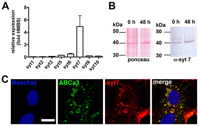
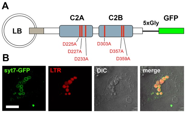
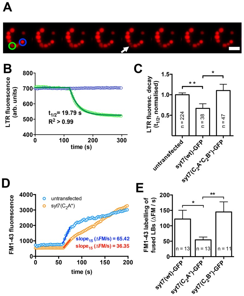
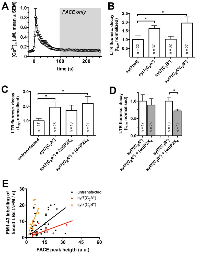
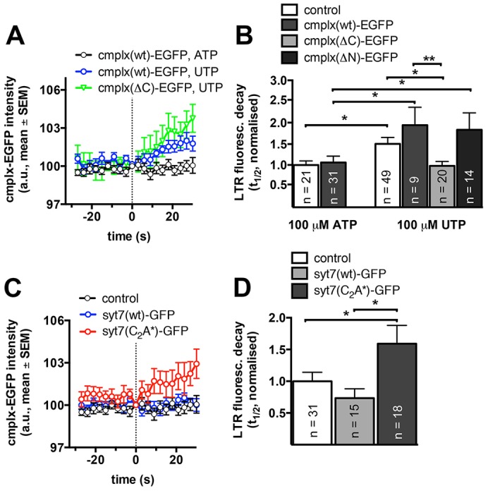
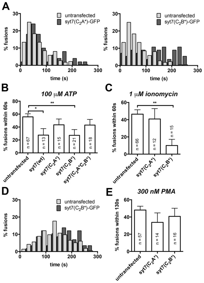
Ca(2+)-dependent regulation of fusion pore dilation and closure is a key mechanism determining the output of cellular secretion. We have recently described 'fusion-activated' Ca(2+) entry (FACE) following exocytosis of lamellar bodies in alveolar type II cells. FACE regulates fusion pore expansion and facilitates secretion. However, the mechanisms linking this locally restricted Ca(2+) signal and fusion pore expansion were still elusive. Here, we demonstrate that synaptotagmin-7 (Syt7) is expressed on lamellar bodies and links FACE and fusion pore dilation. We directly assessed dynamic changes in fusion pore diameters by analysing diffusion of fluorophores across fusion pores. Expressing wild-type Syt7 or a mutant Syt7 with impaired Ca(2+)-binding to the C2 domains revealed that binding of Ca(2+) to the C2A domain facilitates FACE-induced pore dilation, probably by inhibiting translocation of complexin-2 to fused vesicles. However, the C2A domain hampered Ca(2+)-dependent exocytosis of lamellar bodies. These findings support the hypothesis that Syt7 modulates fusion pore expansion in large secretory organelles and extend our picture that lamellar bodies contain the necessary molecular inventory to facilitate secretion during the exocytic post-fusion phase. Moreover, regulating Syt7 levels on lamellar bodies appears to be essential in order that exocytosis is not impeded during the pre-fusion phase.

Keywords: Calcium; Exocytosis; FACE; Fusion pore; Lamellar body; Synaptotagmin.


Figures

Fig. 1.
Fig. 1.
Syt7 is expressed in isolated ATII cells and localised on the lamellar body membrane. (A) Real-time RT-PCR analysis of synaptotagmin transcripts in freshly isolated ATII cells from rat. Data are expressed as the relative expression compared to housekeeping gene Hmbs. Values are mean±s.e.m. from three individual cell isolations and are represented as mean±s.e.m. (B) Western blot of Syt7 from freshly isolated ATII cells and ATII cells 48 h after isolation confirms expression of Syt7 is not altered in cultured cells. Ponceau staining of blots (left) was used to control for equal loading of lanes. (C) Syt7 (red) is primarily localised on lamellar body membranes as detected by indirect immunofluorescence and confirmed by colocalisation with P180 lamellar body protein (green, ABCa3). Scale bar: 10 µm.
Fig. 2.
Fig. 2.
Syt7–GFP localises to the membrane of lamellar bodies. (A) Schematic representation of the Syt7–GFP constructs used in this study. Syt7 and GFP are separated by a short glycine linker. Red lines indicate amino acid positions in the Ca2+-binding sites in the two C2 domains (C2A and C2B) that were mutated to obtain Syt7 constructs with altered Ca2+-binding properties. (B) Syt7–EGFP (green) expressed for 24 h in ATII cells is primarily localised on the limiting membrane of lamellar bodies, as confirmed by co-staining of lamellar bodies with LTR (red). Scale bar: 5 µm.
Fig. 3.
Fig. 3.
Syt7 facilitates fusion pore expansion following exocytic fusion of lamellar bodies with the plasma membrane. (A) Image sequence illustrating loss of LTR from an individual lamellar body following fusion with the plasma membrane and fusion pore opening (green circle). Note that the LTR fluorescence of a non-fusing vesicle (blue circle) does not change significantly. Scale bar: 5 µm. (B) Half-times (t1/2) of the fluorescence decay were analysed to compare diffusion of LTR across the fusion pore for various experimental conditions and to identify differences in fusion pore opening. To analyse t1/2 of the fluorescence decrease upon fusion pore opening the fluorescence of fusing vesicles (green circle in A) was normalized to that of non-fusing vesicles (blue circle in A) to compensate for bleaching, and the decrease of fluorescence was fitted to a one-phase decay. (C) Expression of Syt7(wt)–GFP significantly (P = 0.006) increases the speed of LTR diffusion from fused vesicles indicating faster fusion pore expansion. However, diffusion of LTR from fused lamellar bodies in cells expressing Syt7–GFP that is deficient in Ca2+-binding to the C2A and C2B domain [Syt7(C2A*C2B*)–GFP] was not different to wild-type cells and was significantly (P = 0.03) slower than in cells expressing Syt7(wt)–GFP. (D) Following lamellar body fusion, FM1-43 fluorescence increases owing to incorporation of the dye into the lipidic vesicle contents (Haller et al., 1998). The initial slope of the FM1-43 fluorescence increase (15 s after fusion) was analysed as a direct measure of FM1-43 diffusion across the fusion pore following lamellar body fusion. (E) Diffusion of FM1-43 into fused lamellar bodies was significantly slower in cells expressing a Syt7 mutant deficient in Ca2+-binding to the C2A domain [Syt7(C2A*)–GFP] when compared to cells expressing Syt7(wt)–GFP or Syt7(C2B*)–GFP. Results are mean±s.e.m. *P<0.05; **P<0.01.
Fig. 4.
Fig. 4.
Ca2+ binding to the C2A domain of Syt7 facilitates FACE dependent fusion pore expansion and surfactant secretion. (A) Stimulation of ATII cells with ATP (t = 0), results in a transient rise of the intracellular Ca2+ concentration. To delineate the impact of FACE from the effect of the global rise in Ca2+ on Ca2+-dependent effects of Syt7 on fusion pore expansion, only fusions occurring >100 s following stimulation were analysed (when the global Ca2+ peak has ceased). n = 23 cells. (B) Deletion of Ca2+-binding to the C2A domain of Syt7 [Syt7(C2A*)–GFP or Syt7(C2A*C2B*)–GFP] significantly increased the halftimes of LTR fluorescence decay when compared to Syt7(wt). Expression of Syt7(C2B*)–GFP (intact C2A domain) did not affect LTR fluorescence decay when compared to cells expressing Syt7(wt)–GFP. (C) Modulating the amplitude of FACE by overexpressing either wild-type P2X4 [(wt)P2X4] or (dn)P2X4 (Miklavc et al., 2011) did not affect half-times of LTR fluorescence decay in cells expressing Syt7(C2A*)–GFP. (D) In contrast to cells expressing Syt7(C2A*)–GFP increasing the amplitude of FACE in cells expressing Syt7(C2B*)–GFP significantly increased the speed of LTR diffusion from fused lamellar bodies. (E) The rate of FM1-43 diffusion into fused vesicles at given amplitudes of FACE is increased in cells expressing a functional Syt7 C2A domain [Syt7(C2B*)] and decreased in cells expressing Syt7 with impaired Ca2+ binding to its C2A domain [Syt7(C2A*)] when compared to wild-type cells. Results are mean±s.e.m. *P<0.05.
Fig. 5.
Fig. 5.
FACE and Ca2+ binding to the C2A domain of Syt7 antagonise complexin-2 recruitment to fused lamellar bodies and thereby facilitate fusion pore expansion. (A) Mean fluorescence of cmplx(wt)–GFP and cmplx(ΔC)–GFP before and after lamellar body fusion with the plasma membrane in cells stimulated with either 100 µM ATP (FACE) or 100 µM UTP (no FACE). FACE inhibits increase of cmplx(wt)–GFP at fused lamellar bodies. Fluorescence change was measured in a peri-vesicular region of interest surrounding individual lamellar bodies. The dotted line indicates the time of fusion (data represent a minimum of ten fusions for each condition). (B) Halftimes of LTR fluorescence decay are not significantly different in cells overexpressing cmplx(wt)–GFP when stimulated with 100 µM ATP (FACE). Stimulation with 100 µM UTP (no FACE) resulted in a significant increase in halftimes of LTR fluorescence decay, which was moderately enhanced in cells overexpressing cmplx(wt)–GFP or cmplx(ΔN)–GFP. In contrast, overexpression of cmplx(ΔC)–GFP completely abolished the increase in the halftime of LTR fluorescence decay following stimulation with 100 µM UTP. (C) Mean fluorescence of cmplx(wt)–GFP before and after lamellar body fusion with the plasma membrane in cells stimulated with 100 µM ATP (FACE). Translocation of cmplx(wt)–GFP to fused lamellar bodies is inhibited in wild-type cells (control, black) and cells expressing Syt7(wt)–GFP (blue) but not in cells expressing Syt7(C2A*)–GFP (red). The dotted line indicates the time of fusion (data represent a minimum of ten fusions for each condition). (D) Halftimes of LTR fluorescence decay in cells overexpressing cmplx(wt)-GFP following stimulation with 100 µM ATP. Deletion of Ca2+-binding to the C2A domain of Syt7 [Syt7(C2A*)–GFP] significantly increased the halftimes of LTR fluorescence decay when compared to wild-type cells and cells expressing Syt7(wt)–GFP. Results are mean±s.e.m. *P<0.05.
Fig. 6.
Fig. 6.
The C2A domain of Syt7 impairs Ca2+-dependent lamellar body exocytosis in ATII cells. (A) Expression of Syt7(C2B*)–GFP (right), but not Syt7(C2A*)–GFP (left) resulted in a significant left shift in the fusion delay histograms compared to untransfected cells following stimulation with 100 µM ATP. (B) The percentage of fusions occurring within 60 s of stimulation with 100 µM ATP (during the transient rise in intracellular Ca2+, see Fig. 4A) was significantly reduced in cells expressing Syt7(wt)–GFP or Syt7(C2B*)–GFP compared to untransfected cells. Transfection of ATII cells with Syt7 mutants with impaired Ca2+ binding to the C2A domain [Syt7(C2A*)–GFP or Syt7(C2A*C2B*)–GFP] did not impact on Ca2+-dependent fusion activity. (C) Following stimulation with 1 µM ionomycin, a Ca2+ ionophore that results in a strong long-lasting elevation of the cytoplasmic Ca2+, the effect of Syt7(C2B*)–GFP on inhibiting lamellar body exocytosis was even more significant than following stimulation with ATP. Expression of Syt7(C2A*)–GFP had no significant impact on fusion activity within 60 s of stimulation. (D) When lamellar body exocytosis was stimulated with 300 nM PMA, which does not result in any significant increase in the cytoplasmic Ca2+ concentration (supplementary material Fig. S2) expressing Syt7(C2B*)–GFP did not alter fusion kinetics. (E) No significant difference in the percentage of fusions occurring within 130 s of stimulation with PMA (when ∼50% of fusion occurred in untransfected control cells) were observed between untransfected cells and cells expressing either Syt7(C2A*)–GFP or Syt7(C2B*)–GFP. Results are mean±s.e.m. *P<0.05; **P<0.01.

Similar articles

Cited by

References

    1. Albrecht S., Usmani S. M., Dietl P., Wittekindt O. H. (2010). Plasma membrane trafficking in alveolar type II cells. Cell. Physiol. Biochem. 25, 81–90 10.1159/000272053 - DOI - PubMed
    1. Alvarez de Toledo G., Fernández-Chacón R., Fernández J. M. (1993). Release of secretory products during transient vesicle fusion. Nature 363, 554–558 10.1038/363554a0 - DOI - PubMed
    1. An S. J., Grabner C. P., Zenisek D. (2010). Real-time visualization of complexin during single exocytic events. Nat. Neurosci. 13, 577–583 10.1038/nn.2532 - DOI - PMC - PubMed
    1. Anantharam A., Bittner M. A., Aikman R. L., Stuenkel E. L., Schmid S. L., Axelrod D., Holz R. W. (2011). A new role for the dynamin GTPase in the regulation of fusion pore expansion. Mol. Biol. Cell 22, 1907–1918 10.1091/mbc.E11-02-0101 - DOI - PMC - PubMed
    1. Bhalla A., Tucker W. C., Chapman E. R. (2005). Synaptotagmin isoforms couple distinct ranges of Ca2+, Ba2+, and Sr2+ concentration to SNARE-mediated membrane fusion. Mol. Biol. Cell 16, 4755–4764 10.1091/mbc.E05-04-0277 - DOI - PMC - PubMed

Publication types

MeSH terms