. 2015 May;43(5):1071-88.
doi: 10.1007/s10439-014-1171-9. Epub 2014 Oct 25.

Head rotational acceleration characteristics influence behavioral and diffusion tensor imaging outcomes following concussion

Affiliations

Head rotational acceleration characteristics influence behavioral and diffusion tensor imaging outcomes following concussion

Brian D Stemper et al. Ann Biomed Eng. 2015 May.

Abstract

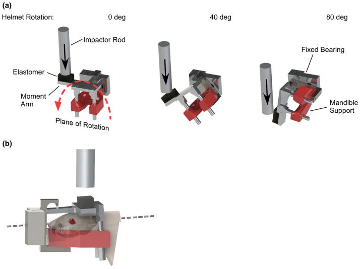
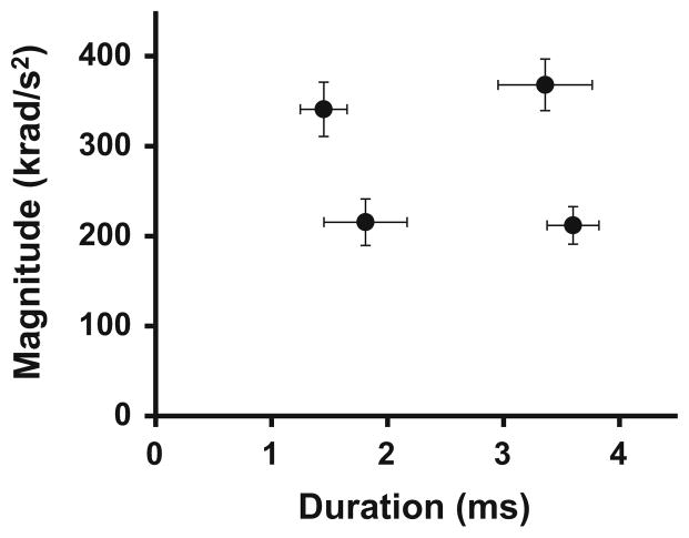
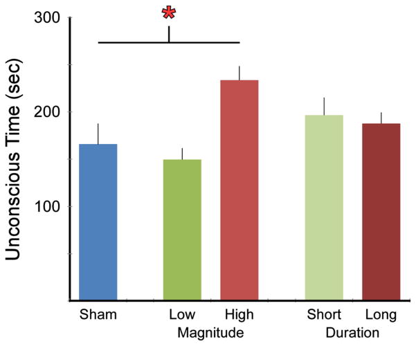
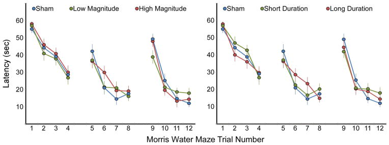
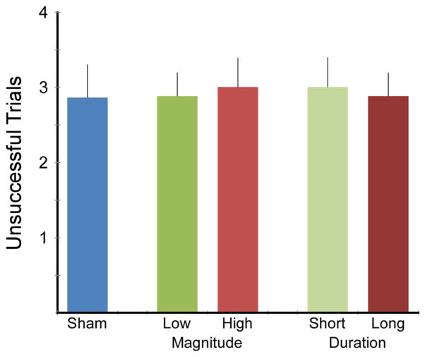
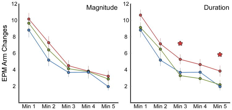
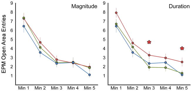
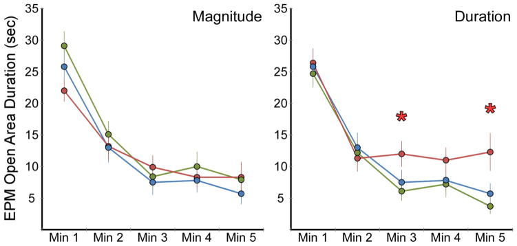
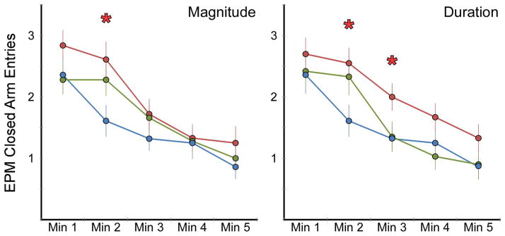
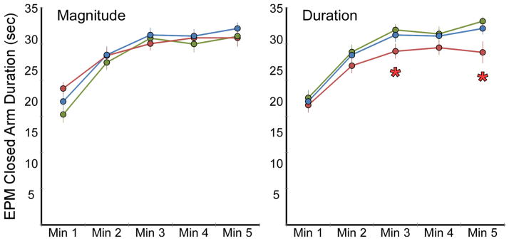
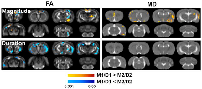
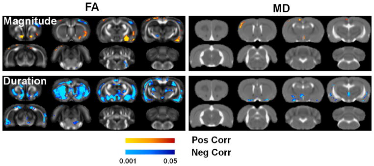
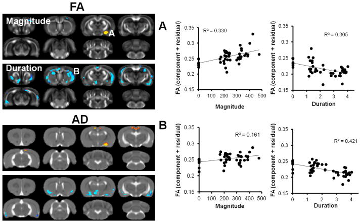
A majority of traumatic brain injuries (TBI) in motor vehicle crashes and sporting environments are mild and caused by high-rate acceleration of the head. For injuries caused by rotational acceleration, both magnitude and duration of the acceleration pulse were shown to influence injury outcomes. This study incorporated a unique rodent model of rotational acceleration-induced mild TBI (mTBI) to quantify independent effects of magnitude and duration on behavioral and neuroimaging outcomes. Ninety-two Sprague-Dawley rats were exposed to head rotational acceleration at peak magnitudes of 214 or 350 krad/s(2) and acceleration pulse durations of 1.6 or 3.4 ms in a full factorial design. Rats underwent a series of behavioral tests including the Composite Neuroscore (CN), Elevated Plus Maze (EPM), and Morris Water Maze (MWM). Ex vivo diffusion tensor imaging (DTI) of the fixed brains was conducted to assess the effects of rotational injury on brain microstructure as revealed by the parameter fractional anisotropy (FA). While the injury did not cause significant locomotor or cognitive deficits measured with the CN and MWM, respectively, a main effect of duration was consistently observed for the EPM. Increased duration caused significantly greater activity and exploratory behaviors measured as open arm time and number of arm changes. DTI demonstrated significant effects of both magnitude and duration, with the FA of the amygdala related to both the magnitude and duration. Increased duration also caused FA changes at the interface of gray and white matter. Collectively, the findings demonstrate that the consequences of rotational acceleration mTBI were more closely associated with duration of the rotational acceleration impulse, which is often neglected as an independent factor, and highlight the need for animal models of TBI with strong biomechanical foundations to associate behavioral outcomes with brain microstructure.


Figures

FIGURE 1
FIGURE 1
(a) MCW rotational injury device that was used to produce mild TBI in rats through coronal plane rotational acceleration of the head. The three images demonstrate helmet positioning just prior to impact from the rod (left), and during impact causing the helmet to rotate 40° (middle) and 80° (right). Total rotation of the helmet was limited to 90°. Motion was constrained to pure coronal plane rotation, without linear displacement, using a fixed bearing located anterior to the helmet. (b) The axis of helmet rotation was located in the mid-sagittal plane in the lower half of the brain.
FIGURE 2
FIGURE 2
Mean and standard deviation values for the four experimental groups focused on two levels of rotational acceleration magnitude (M1 and M2) and two levels of rotational acceleration duration (D1 and D2). Biomechanics for each group were distinct and demonstrated reasonable repeatability, with coefficient of variation of 10.5 and 16.5% for magnitude and duration, respectively.
FIGURE 3
FIGURE 3
Duration of unconsciousness following exposure to head rotational acceleration was significantly dependent (p<0.0001) upon acceleration magnitude but not duration, wherein rats exposed to high magnitude accelerations remained unconscious for a significantly longer time than shams or rats exposed to low magnitude accelerations.
FIGURE 4
FIGURE 4
Rats were subjected to the Morris Water Maze Visuo-Spatial Learning Paradigm on the first, second, and third days post injury. One set per day consisted of four trials with the platform located in the same location, but moved between days. No statistically significant differences in latency as an effect of increasing magnitude or duration were evident.
FIGURE 5
FIGURE 5
Total number of unsuccessful trials across all three sets of the Morris Water Maze assessment. Unsuccessful trials that occurred in the first trial of each set were not included because the platform was moved to a new location for each set. The number of unsuccessful trials was not different between shams and any combination of injured groups, indicating a lack of any significant spatial learning deficits.
FIGURE 6
FIGURE 6
Rats were exposed to the Elevated Plus Maze for 5 min on the second day post injury. Total number of arm changes between closed arms, the center platform, and open arms were counted and used as an assessment of post-injury activity. Results for magnitude/duration are presented as high magnitude/long duration (red), low magnitude/short duration (green), and shams (blue). The number of arm changes was significantly dependent upon duration (p<0.05) but not magnitude of rotational acceleration. Independent minute-by-minute analyses revealed significantly increased activity (p<0.05) in the long duration group during the third and fifth minutes of the assessment.
FIGURE 7
FIGURE 7
Rats were exposed to the Elevated Plus Maze for 5 min on the second day post injury. Total number of open area entries (center platform and open arms) were counted and used as an assessment of post-injury activity and inhibition. Results for magnitude/duration are presented as high magnitude/long duration (red), low magnitude/short duration (green), and shams (blue). The number of open area entries was significantly dependent upon duration (p<0.05) but not magnitude of rotational acceleration. Independent minute-by-minute analyses revealed significantly increased entries (p<0.05) in the long duration group during the third and fifth minutes of the assessment.
FIGURE 8
FIGURE 8
Rats were exposed to the Elevated Plus Maze for 5 min on the second day post injury. Total amount of time spent in the open areas (center platform and open arms) was used as an assessment of post-injury inhibition. Results for magnitude/ duration are presented as high magnitude/long duration (red), low magnitude/short duration (green), and shams (blue). The total amount of time spent in open areas was significantly dependent upon duration (p<0.05) but not magnitude of rotational acceleration. Independent minute-by-minute analyses revealed significantly increased open area time (p <0.05) in the long duration group during the third and fifth minutes of the assessment.
FIGURE 9
FIGURE 9
Rats were exposed to the Elevated Plus Maze for 5 min on the second day post injury. Total number of closed arm entries were counted and used as an assessment of post-injury activity and inhibition. Results for magnitude/duration are presented as high magnitude/long duration (red), low magnitude/short duration (green), and shams (blue). The number of closed arm entries was significantly dependent upon duration (p <0.05) and magnitude (p <0.05) of rotational acceleration. Independent minute-by-minute analyses revealed significantly increased entries (p <0.05) in the long duration group during the third and fifth minutes of the assessment.
FIGURE 10
FIGURE 10
Rats were exposed to the Elevated Plus Maze for 5 min on the second day post injury. Total amount of time spent in the closed arms was used as an assessment of post-injury inhibition. Results for magnitude/duration are presented as high magnitude/long duration (red), low magnitude/short duration (green), and shams (blue). The total amount of time spent in the closed arms was significantly dependent upon duration (p<0.05) but not magnitude of rotational acceleration. Independent minute-by-minute analyses revealed significantly decreased closed arm time (p <0.05) in the long duration group during the third and fifth minutes of the assessment.
FIGURE 11
FIGURE 11
DTI group-wise factorial analysis. Voxels with a significant main effect of either magnitude or duration solely within the animals exposed to injury. Regions of decreased FA in the high magnitude group were evident in portions of the corticospinal tracts and internal capsule, whereas FA was increased in the high magnitude group in the amygdala. MD was increased in the high magnitude group predominantly in portions of the right ventral cortex and white matter. Regions of decreased FA were evident in the high duration groups, predominantly located bilaterally at the interface of gray and white matter in ventral regions. MD did not exhibit any effects of duration. Only voxels with a significant main effect (p <0.05 and cluster size >200 voxels) for either magnitude or duration in an F-test are shown, with the colors reflecting the p-value and sign of the post hoc T-statistic.
FIGURE 12
FIGURE 12
DTI regression analysis. Voxels with a significant linear regression with the magnitude or duration measured from each injury are shown for FA and MD. Increasing magnitude was associated with increased FA in the right amygdala and ventral white matter, whereas decreased FA was evident in the right dorsolateral cortex. Increasing duration was significantly associated with a much larger portion of the brain, localized bilaterally in the ventral cortex and white matter and large portions of the basal ganglia. Only few small regions were significant in the regression analysis between MD and either magnitude or duration. The colored regions depict the p value of the T-statistic for either magnitude or duration, while controlling for the other parameter, with a p value threshold of 0.05 (two-tailed) and cluster size threshold of 200 voxels.
FIGURE 13
FIGURE 13
DTI conjunction analysis. Regions having both a significant main effect and a significant correlation with either magnitude or duration are shown. The amygdala is the most pronounced region exhibiting a strong effect of magnitude. The average FA from the significant voxels was derived from each animal, and the partial regression plots are shown in (a). For duration, the ventral portion of the gray/white matter interface was highly significant, and the mean FA from this region is shown in the partial regression plots in (b). The conjunction analysis revealed most of the FA changes were associated with changes in AD. Other DTI parameters (MD & RD) did not have any significant voxels in the conjunction analysis.

Similar articles

Cited by

References

    1. Abdel Baki SG, Kao HY, Kelemen E, et al. A hierarchy of neurobehavioral tasks discriminates between mild and moderate brain injury in rats. Brain Res. 2009;1280:98–106. - PubMed
    1. Abel JM, Gennarelli TA, Segawa H. Incidence and severity of cerebral concussion in the rhesus monkey following sagittal plane angular acceleration. 22nd Stapp Car Crash Conference; Ann Arbor, MI. 1978. pp. 35–53.
    1. Bao F, Shultz SR, Hepburn JD, et al. A CD11d monoclonal antibody treatment reduces tissue injury and improves neurological outcome after fluid percussion brain injury in rats. J Neurotrauma. 2012;29:2375–2392. - PMC - PubMed
    1. Baykara B, Cetin F, Baykara B, et al. Anxiety caused by traumatic brain injury correlates to decreased prefrontal cortex VEGF immunoreactivity and neuron density in immature rats. Turk Neurosurg. 2012;22:604–610. - PubMed
    1. Bolouri H, Saljo A, Viano DC, et al. Animal model for sport-related concussion; ICP and cognitive function. Acta Neurol Scand. 2012;125:241–247. - PubMed

Publication types