. 2014 Dec 1;104(3):501-11.
doi: 10.1093/cvr/cvu231. Epub 2014 Oct 24.

LQT1 mutations in KCNQ1 C-terminus assembly domain suppress IKs using different mechanisms

Affiliations

LQT1 mutations in KCNQ1 C-terminus assembly domain suppress IKs using different mechanisms

Ademuyiwa S Aromolaran et al. Cardiovasc Res. .

Abstract

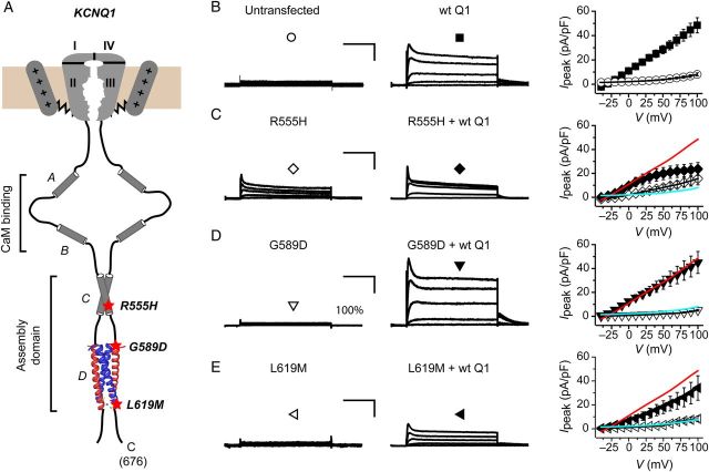
Aims: Long QT syndrome 1 (LQT1) mutations in KCNQ1 that decrease cardiac IKs (slowly activating delayed rectifier K(+) current) underlie ventricular arrhythmias and sudden death. LQT1 mutations may suppress IKs by preventing KCNQ1 assembly, disrupting surface trafficking, or inhibiting gating. We investigated mechanisms underlying how three LQT1 mutations in KCNQ1 C-terminus assembly domain (R555H/G589D/L619M) decrease IKs in heterologous cells and cardiomyocytes.

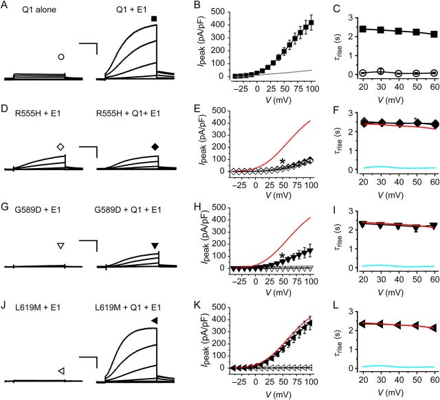
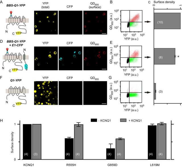
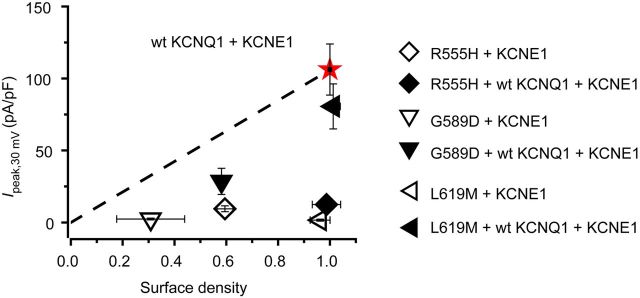
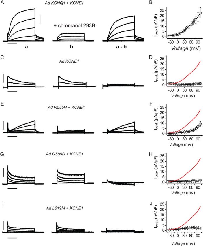
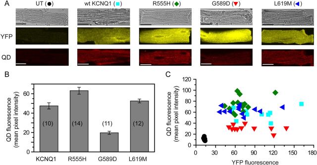
Methods and results: In Chinese hamster ovary (CHO) cells, mutant KCNQ1 + KCNE1 channels either produced no currents (G589D/L619M) or displayed markedly reduced IKs with a right-shifted voltage-dependence of activation (R555H). When co-expressed with wild-type (wt) KCNQ1, the mutant KCNQ1s displayed varying intrinsic dominant-negative capacities that were affected by auxiliary KCNE1. All three mutant KCNQ1s assembled with wt KCNQ1 as determined by fluorescence resonance energy transfer (FRET). We developed an optical quantum dot labelling assay to measure channel surface density. G589D/R555H displayed substantial reductions in surface density, which were either partially (G589D) or fully (R555H) rescued by wt KCNQ1. Unexpectedly, L619M showed no trafficking defect. In adult rat cardiomyocytes, adenovirus-expressed homotetrameric G589D/L619M + KCNE1 channels yielded no currents, whereas R555H + KCNE1 produced diminished IKs with a right-shifted voltage-dependence of activation, mimicking observations in CHO cells. In contrast to heterologous cells, homotetrameric R555H channels showed no trafficking defect in cardiomyocytes.

Conclusion: Distinct LQT1 mutations in KCNQ1 assembly domain decrease IKs using unique combinations of biophysical and trafficking mechanisms. Functional deficits in IKs observed in heterologous cells are mostly, but not completely, recapitulated in adult rat cardiomyocytes. A 'methodological chain' combining approaches in heterologous cells and cardiomyocytes provides mechanistic insights that may help advance personalized therapy for LQT1 mutations.

Keywords: Cardiac myocyte; Channel trafficking; Ion channel; KCNQ1; Long QT syndrome.


Figures

Figure 1
Figure 1
Electrophysiological properties of wt and mutant KCNQ1 in the absence of KCNE1. (A) Schematic showing the modular structure of KCNQ1 C-terminus and relative locations of LQT1 mutations evaluated in this study. The structure of the coiled-coil tetramerization domain (helix D) is reproduced from PDB ID: 3BJ4. (B) Representative traces from CHO cells either untransfected (left panel) or expressing wt KCNQ1-YFP (middle). (Right panel) Population Ipeak–V curves for untransfected (open circle) or wt KCNQ1-expressing (filled square, n = 47) cells. (C) Exemplar currents from CHO cells expressing R555H-YFP alone (left) or with wt KCNQ1-YFP (middle). (Right panel) Population Ipeak–V curves for R555H expressed alone (open diamond, n = 12) or with wt KCNQ1 (filled diamond, n = 5). IpeakV curve for wt KCNQ1-YFP is reproduced (red line) to facilitate visual comparison. (D and E) Exemplar currents and IpeakV relationships for indicated KCNQ1 mutants, same format as C. Data points are means ± SEM, n = 6–10. Scale bars, 10 pA/pF, 1 s. Displayed exemplar currents were obtained from test pulses to −40, −10, +10, +40, +70, and +100 mV for all experimental conditions.
Figure 2
Figure 2
Electrophysiological properties of wt and mutant KCNQ1 in the presence of KCNE1. (A) Exemplar currents from CHO cells expressing KCNQ1 alone (left) or KCNQ1-YFP + KCNE1 (right). (B) Population Ipeak–V curves for KCNQ1-YFP + KCNE1 channels (filled square, n = 11). Data for KCNQ1 alone channels are reproduced from Figure 1B (grey trace). (C) Voltage-dependence of time constants for activation (τrise) of wt KCNQ1 alone (open circle, n = 47) and wt KCNQ1 + KCNE1 (filled square, n = 11) currents. (D) Exemplar currents from CHO cells expressing (left) R555H + KCNE1 (homotetrameric) or (right) R555H + wt KCNQ1 + KCNE1 (heterotetrameric). (E) Population Ipeak–V curves for homotetrameric (open diamond, n = 18) and heterotetrameric (filled diamond, n = 7) R555H channels. Data for control wt KCNQ1 + KCNE1 channels are reproduced (red trace) for comparison. (F) Voltage-dependence of τrise for homotetrameric (open diamond, n = 18) and heterotetrameric (filled diamond, n = 7) R555H channels. Data for wt KCNQ1 alone (cyan trace) and wt KCNQ1 + KCNE1 (red trace) channels are shown for comparison. (G–L) Exemplar currents, IpeakV relationships, and τrise–V curves for homotetrameric and heterotetrameric channels containing the indicated KCNQ1 mutants, same format as D–F. Data points are mean ± SEM, n = 5–21. Scale bars, 20 pA/pF, 1 s. Displayed exemplar currents were obtained from test pulses to −40, −10, +10, +40, +70, and +100 mV for all experimental conditions.
Figure 3
Figure 3
Optical detection of cell surface BBS-tagged KCNQ1 with quantum dot. (A, left) Schematic showing quantum dot (QD655) labelling of cell surface BBS-tagged KCNQ1-YFP. (Middle) Confocal images of non-permeabilized cells expressing BBS-KCNQ1-YFP labelled with QD655. Panels show channels for YFP (total expression), CFP, and QD655 (surface channels). Scale bar, 20 µm. (B) Flow cytometry dot plot showing QD655 vs. YFP fluorescence intensity signals for cells expressing BBS-KCNQ1-YFP. About 50 000 cells were counted. Vertical and horizontal lines represent threshold values set based on isochronal untransfected cells. Each dot represents a single cell. Dots have been arbitrarily coloured to facilitate visualization of distinct populations. Green dots represent BBS-KCNQ1-YFP-expressing cells with little QD655 signal, indicating low channel surface density. Red dots denote BBS-KCNQ1-YFP-positive cells with robust channel trafficking to the surface. Black dots in bottom left quadrant are untransfected cells. (C) Normalized mean QD655 signals from YFP-positive cells provide an index of relative surface density. (D and E) Data for cells expressing BBS-KCNQ1-YFP + KCNE1-CFP, same format as A–C. (F and G) Negative control. Data for cells expressing untagged KCNQ1-YFP, same format as A–C. *P < 0.05 compared with BBS-KCNQ1-YFP, unpaired Student's t-test (scale bars, 20 μm). (H) Computed relative surface density for BBS-tagged wt and LQT1 mutant KCNQ1-YFP subunits in the absence and presence of untagged wt KCNQ1.
Figure 4
Figure 4
Relative contributions of biophysical and trafficking mechanisms to IKs suppression for distinct LQT1 mutations. Plot of peak IKs recorded at +30 mV (Ipeak, 30mV) vs. relative surface density for wt KCNQ1 + KCNE1 (star) and indicated mutant KCNQ1 + KCNE1 ± wt KCNQ1. Dotted line from origin to wt KCNQ1 data point tracks expected relationship between Ipeak,+30 mV and relative surface density if mutations affected only channel trafficking with no biophysical effects. Deviations from this line report on biophysical mechanisms contributing to observed changes in IKs.
Figure 5
Figure 5
Reconstitution of wt and mutant IKs in adult rat ventricular cardiomyocytes. (A) Exemplar current traces measured in the absence (traces a) and presence (traces b) of 30 µM chromanol 293B in a cardiomyocyte expressing wt KCNQ1 + KCNE1. The difference traces (a–b) represent the chromanol 293B-sensitive currents, i.e. IKs. (B) Population I–V curves for chromanol 293B-sensitive currents in adult rat cardiomyocytes expressing wt KCNQ1 + KCNE1, n = 26. (C and D) Data for cardiomyocytes expressing KCNE1 alone indicate the lack of IKs (n = 4), same format as A and B. (E–J) Data obtained from cardiomyocytes expressing R555H + KCNE1 (n = 21), G589D + KCNE1 (n = 10), and L619M + KCNE1 (n = 16), respectively, same format as A and B. Data for wt KCNQ1 + KCNE1 channels are reproduced (red trace) in E–J for visual comparison. Data points are mean ± SEM. Data were generated from three different rat cardiomyocyte preparations. Scale bars, 5 pA/pF, 1 s.
Figure 6
Figure 6
Detection of surface KCNQ1 channels with quantum dot in cardiomyocytes. (A) Confocal images of non-permeabilized adult rat ventricular cardiomycytes showing bright-field, YFP, and QD655 fluorescence images. Panels show uninfected control myocytes and cardiomyocytes expressing wt and mutant BBS-tagged KCNQ1 subunits. Scale bars, 20 µm. (B) QD655 fluorescence mean pixel intensity for the different conditions. (C) Scatter plots of QD655 (surface channels) vs. YFP fluorescence (total expression) for the different BBS-tagged wt (filled square) and mutant KCNQ1-YFP subunits (R555H, filled diamond; G589D, inverted triangle; L619M, left-pointing filled triangle). Data were generated from two different rat cardiomyocyte preparations.

Similar articles

Cited by

References

    1. Nerbonne JM, Kass RS. Molecular physiology of cardiac repolarization. Physiol Rev. 2005;85:1205–1253. - PubMed
    1. Charpentier F, Merot J, Loussouarn G, Baro I. Delayed rectifier K(+) currents and cardiac repolarization. J Mol Cell Cardiol. 2010;48:37–44. - PubMed
    1. Sanguinetti MC, Curran ME, Zou A, Shen J, Spector PS, Atkinson DL, Keating MT. Coassembly of K(V)LQT1 and minK (IsK) proteins to form cardiac I(Ks) potassium channel. Nature. 1996;384:80–83. - PubMed
    1. Barhanin J, Lesage F, Guillemare E, Fink M, Lazdunski M, Romey G. K(V)LQT1 and lsK (minK) proteins associate to form the I(Ks) cardiac potassium current. Nature. 1996;384:78–80. - PubMed
    1. Takumi T, Ohkubo H, Nakanishi S. Cloning of a membrane protein that induces a slow voltage-gated potassium current. Science. 1988;242:1042–1045. - PubMed

Publication types