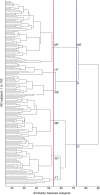
Anatomical heterogeneity of Alzheimer disease: based on cortical thickness on MRIs
- PMID: 25344382
- PMCID: PMC4248459
- DOI: 10.1212/WNL.0000000000001003
Anatomical heterogeneity of Alzheimer disease: based on cortical thickness on MRIs
Abstract
Objective: Because the signs associated with dementia due to Alzheimer disease (AD) can be heterogeneous, the goal of this study was to use 3-dimensional MRI to examine the various patterns of cortical atrophy that can be associated with dementia of AD type, and to investigate whether AD dementia can be categorized into anatomical subtypes.
Methods: High-resolution T1-weighted volumetric MRIs were taken of 152 patients in their earlier stages of AD dementia. The images were processed to measure cortical thickness, and hierarchical agglomerative cluster analysis was performed using Ward's clustering linkage. The identified clusters of patients were compared with an age- and sex-matched control group using a general linear model.
Results: There were several distinct patterns of cortical atrophy and the number of patterns varied according to the level of cluster analyses. At the 3-cluster level, patients were divided into (1) bilateral medial temporal-dominant atrophy subtype (n = 52, ∼ 34.2%), (2) parietal-dominant subtype (n = 28, ∼ 18.4%) in which the bilateral parietal lobes, the precuneus, along with bilateral dorsolateral frontal lobes, were atrophic, and (3) diffuse atrophy subtype (n = 72, ∼ 47.4%) in which nearly all association cortices revealed atrophy. These 3 subtypes also differed in their demographic and clinical features.
Conclusions: This cluster analysis of cortical thickness of the entire brain showed that AD dementia in the earlier stages can be categorized into various anatomical subtypes, with distinct clinical features.
© 2014 American Academy of Neurology.
Conflict of interest statement
The authors report no disclosures relevant to the manuscript. Go to
Figures


References
-
- Kramer JH, Miller BL. Alzheimer's disease and its focal variants. Semin Neurol 2000;20:447–454. - PubMed
-
- Johnson JK, Head E, Kim R, Starr A, Cotman CW. Clinical and pathological evidence for a frontal variant of Alzheimer disease. Arch Neurol 1999;56:1233–1239. - PubMed
-
- Butters MA, Lopez OL, Becker JT. Focal temporal lobe dysfunction in probable Alzheimer's disease predicts a slow rate of cognitive decline. Neurology 1996;46:687–692. - PubMed
-
- Tang-Wai DF, Graff-Radford NR, Boeve BF, et al. . Clinical, genetic, and neuropathologic characteristics of posterior cortical atrophy. Neurology 2004;63:1168–1174. - PubMed
Publication types
MeSH terms
LinkOut - more resources
Full Text Sources
Other Literature Sources
Medical
