

Cavernous hemangioma of thymus misdiagnosed as thymoma: a case report
- PMID: 25344424
- PMCID: PMC4219033
- DOI: 10.1186/1477-7819-12-323
Cavernous hemangioma of thymus misdiagnosed as thymoma: a case report
Abstract
Introduction: Cavernous hemangioma in the thymus is a rare presentation in mediastinal hemangiomas. The diagnosis is difficult to make promptly because both invasive and noninvasive examinations usually fail to distinguish it from other tumors of the mediastinum. Their clinical presentations depends on their size and their involvement with adjacent mediastinal structures.
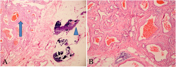
Case presentation: We treated a 52-year-old man with thymic cavernous hemangioma that was incidentally detected by chest radiography during a routine health check, and had been misdiagnosed as thymoma before the operation. The tumor was completely resected by thymectomy via video-assisted thoracic surgery. The pathological tissue was diagnosed as a cavernous hemangioma, and no phlebolith was observed in the center.
Conclusions: We reported this case of thymic cavernous hemangioma for its extremely rare occurrence in the thymus. The preoperative diagnosis remains a challenge both clinically and radiologically. It is still difficult to distinguish this tumor from other tumors in the thymus. Furthermore, biopsies might not result in a definitive diagnosis. Finally, surgical resection provides material for histopathologic diagnosis. To facilitate the preoperative diagnosis of such a rare tumor, more cases will need to be reported.
Figures

References
-
- Romeo GP, Fiedler PN, Hecht CS, Nuñez D. Thrombosed cavernous hemangioma arising in cervical ectopic thymus tissue: a case report. Conn Med. 2012;76:401–404. - PubMed
-
- Wychulis AR, Payne WS, Clagett OT, Woolner LB. Surgical treatment of mediastinal tumors: a 40 year experience. J Thorac Cardiovasc Surg. 1971;62:379–392. - PubMed
-
- Yamazaki A, Miyamoto H, Saito Y, Matsuzawa H, Sakao Y, Anami Y. Cavernous hemangioma of the anterior mediastinum: case report and 50-year review of Japanese cases. Jpn J Thorac Cardiovasc Surg. 2006;54:221–224. - PubMed
-
- Niedzwiecki G, Wood BP. Radiological cases of the month. Thymic hemangioma. Am J Dis Child. 1990;144:1149–1150. - PubMed
Publication types
MeSH terms
LinkOut - more resources
Full Text Sources
Other Literature Sources
Medical
