. 2014 Dec 1;193(11):5534-44.
doi: 10.4049/jimmunol.1401655. Epub 2014 Oct 24.

T cell-B cell thymic cross-talk: maintenance and function of thymic B cells requires cognate CD40-CD40 ligand interaction

Affiliations

T cell-B cell thymic cross-talk: maintenance and function of thymic B cells requires cognate CD40-CD40 ligand interaction

Chiharu Fujihara et al. J Immunol. .

Abstract

Thymic development requires bidirectional interaction or cross-talk between developing T cells and thymic stromal cells, a relationship that has been best characterized for the interaction between thymocytes and thymic epithelial cells. We have characterized in this article the requirement for similar cross-talk in the maintenance and function of thymic B cells, another population that plays a role in selection of developing thymic T cells. We found that maintenance of thymic B cells is strongly dependent on the presence of mature single-positive thymocytes and on the interactions of these T cells with specific Ag ligand. Maintenance of thymic B cell number is strongly dependent on B cell-autonomous expression of CD40, but not MHC class II, indicating that direct engagement of CD40 on thymic B cells is necessary to support their maintenance and proliferation. Thymic B cells can mediate negative selection of superantigen-specific, self-reactive, single-positive thymocytes, and we show that CD40 expression on B cells is critical for this negative selection. Cross-talk with thymic T cells is thus required to support the thymic B cell population through a pathway that requires cell-autonomous expression of CD40, and that reciprocally functions in negative selection of autoreactive T cells.


Figures
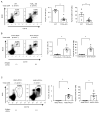
Figure 1
Figure 1
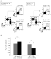
Antigen recognition by SP T cells positively regulates the thymic B cell population. Thymocytes from 10–12 week-old WT and TCRα KO mice, and 6–12 week-old OTII/RIP-OVA transgenic mice and AND/PCC TCR transgenic mice were stained with anti-B220 and anti-CD19 mAbs. Thymic B cell frequency and absolute number are shown. (A) Data are combined from 3 independent experiments. Total numbers of mice: WT: n=6 and TCRα KO: n=8. (B) Data are combined from 4 independent experiments, each using littermate mice. Total numbers of mice: OTII+/RIP-OVA TCR transgenic mice: n=9 and OTII+/RIP-OVA+ TCR transgenic mice: n=7.(C) Data are combined from 5 independent experiments, each using littermate mice. Total numbers of mice: AND+/PCC TCR transgenic mice: n=6 and AND+/PCC+ TCR transgenic mice: n=7. Dot plots: data are representative of the experiments. Bar graph: Mean ± SE. Statistical significance was determined using a Student’s non-paired two-tailed t-test. * P<0.05, ** P<0.01.
Figure 2
Figure 2
Thymic B cells do not require cell-autonomous MHCII expression for their maintenance.(A) Thymocytes from mixed bone marrow chimeras were stained with anti-CD45.1 (WT) and anti-CD45.2 (WT or MHCII KO) Abs to distinguish the congenic donors. Both donor-derived thymocyte populations were further gated on B220+CD19+ thymic B cells. Group 1 (Left) Equal numbers of WT (CD45.1) + MHCII KO (CD45.2) bone marrow cells were used to reconstitute irradiated WT (CD45.1 x CD45.2) recipients; Group 2(Right) Equal numbers of WT (CD45.1) + WT (CD45.2) bone marrow cells were used to reconstitute irradiated WT (CD45.1 x CD45.2) recipients. Data are representative of 9 chimeras. (B) Thymic B cell frequency in each donor-derived population in Group 1 and Group 2. Data from two independent sets of chimeras consisting of 9 mice in each group were combined for this analysis. Statistical significance was determined using a Student’s paired two-tailed t-test. NS, not significant.
Figure 3
Figure 3
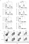
Thymic B cell maintenance is dependent upon CD40L-CD40 interactions.(A) Thymic B cell frequencies in WT, CD80/CD86/CD40 triple KO (T KO) CD80/CD86 double KO (D KO) and CD40 KO mice on a B6 background (Left) and BALB background (Right). (B) Thymic B cell frequencies in WT, CD40 KO mice and CD40L KO mice on B6 background (Left) and BALB background (Right). B6 WT: n=12, B6 T KO: n=6, B6 D KO: n=7, B6 CD40 KO: n=5 and B6 CD40L KO: n=3. Data are combined from 3 independent experiments. BALB WT: n=9, BALB T KO: n=6, BALB D KO: n=7, BALB CD40 KO: n=9 and BALB CD40L KO: n=6. Data are combined from 4 independent experiments. Mean ± SE. Statistical significance was determined using one-way ANOVA. * P<0.05, ** P<0.01, *** P<0.001, **** P<0.0001. (C) Comparison of B cell frequencies between BALB WT and BALB CD40 KO mice in thymus, spleen, lymph nodes (LN) and blood. Data are representative of 3 independent experiments.
Figure 4
Figure 4
CD40 supports proliferation in thymic B cells. CD4 cells were depleted from freshly explanted thymocytes pooled from 2–4 WT and CD40 KO mice using CD4 micro beads. CD4-depleted thymocytes were stained for B cell-surface molecules, B220 and CD19, fixed and permeabilized before intracellular staining. (A) Cell cycle analysis. CD4-depleted thymocytes were stained with DAPI for cell cycle analysis. Panels show DAPI staining profiles gated on B220+CD19+ thymic B cells from WT (Left) and CD40 KO (Right). Data are representative of 3 independent experiments. Graph shows thymic B cell frequency in each phase of cell cycles for WT (Gray) and CD40 KO (Black). Mean ± SE for n=3. Statistical significance was determined using a Student’s non-paired two-tailed t-test. * P<0.05. (B) Cleaved-Caspase-3 expression. Freshly explanted thymic B cells from WT and CD40 KO mice, freshly explanted WT double negative 3 thymocytes (DN3) and overnight incubated WT CD4SP T cells were stained with anti-cleaved-Caspase-3 Ab for apoptosis analysis. Graph showscleaved-Caspase-3 positive frequencies among B220+CD19+ thymic B cells, DN3 cells and CD4 SP T cells incubated overnight. CD4 SP T cells incubated overnight were used as an apoptosis positive control and freshly explanted DN3cells were used as an apoptosis negative control. Mean ± SE for n=3. Statistical significance was determined using a Student’s non-paired two-tailed t-test between WT and CD40 KO. NS, not significant.(C) CD4-depleted thymocytes from WT and CD40 KO mice were stained with anti-Bcl-2 mAb. Histogram shows Bcl-2 profile gated on B220+CD19+ thymic B cells. WT (Thick line) and CD40 KO (Dotted line), isotype control (Gray filled). Data are representative of 3 independent experiments.
Figure 5
Figure 5
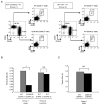
Cell-autonomous CD40 expression is important for maintenance of thymic B cells. Group 1: BM cells from WT (CD45.1) and CD40 KO (CD45.2) were transplanted into irradiated WT (CD45.1 x CD45.2) mice. Group 2: BM cells from WT (CD45.1) and WT (CD45.2) were transplanted into WT (CD45.1 x CD45.2) mice as a control. Chimeras were analyzed for thymic B cell and DC frequencies 8–12 weeks after reconstitution. (A) Thymocytes from recipients were stained with anti-CD45.1 (WT) and anti-CD45.2 (WT or CD40 KO) mAbsto distinguish congenic donors. Each donor-derived thymocyte population was gated on B220+CD19+ thymic B cells or CD11c+ DCs. Group 1: left and Group 2: right. Graphs show (B) thymic B cell frequency in Group 1 and 2 and (C) thymic DC frequency in Group 1. Data from 3 independent sets of chimeras consisting of 13 mice in each group were combined for this analysis. Statistical significance was determined using a Student’s paired two-tailed t-test. ** P<0.01.
Figure 6
Figure 6
CD40 on B cells is essential for superantigen-mediated negative selection of CD4 T cells. (A) Superantigen-mediated negative selection was analyzed in BALB WT, BALB B cell KO, and B6 WT mice. Thymocytes from BALB WT, BALB B cell KO and B6 WT mice were stained with anti-TCRVβ8, Vβ11 and Vβ12mAbs. Graph shows frequency of thymicCD4 SP T cells expressing TCRVβ8, Vβ11 and Vβ12 in BALB WT (Black), BALB B cell KO (Gray) and B6 WT (White). TCRVβ8-expressing CD4 SP T cells are shown as a control not affected by superantigen-specific deletion. Mean ± SE, n=3 per strain. Statistical significance was determined using a Student’s paired two-tailed t-test.** P<0.01.(B) BM cells from BALB B cell KO and BALB CD40 KO were transplanted into irradiated B cell KO mice, generating mice specifically deficient for expression of CD40 on B cells (Group 1). BM cells from BALB B cell KO and BALB WT were transplanted into B cell KO mice as a control (Group 2). Chimeras were studied 8–12 weeks after reconstitution. Frequency of thymic CD4 SP T cells expressing TCRVVβ8, Vβ11 and Vβ12 in Group 1 (B cell KO+CD40 KO BM donors) (Black), Group 2 (B cell KO+WT BM donors) (Gray), and control B6 WT (White). The graph shows mean ± SE for 4 mice per recipient in each group. Statistical significance was determined using a Student’s paired two-tailed t-test.** P<0.01.(C) In vitro negative selection was performed using BALB WT and BALBCD40 KO thymic B cells. B6 CD4+CD8lo DP thymocytes, BALB WT and CD40 KO thymic B cells and B6 and BALB splenic B cells were sorted, and thymocytes and B cells were co-cultured for 24–36h. The TCRVβ11- and TCRVβ12-expressing CD4 SP T cell frequencies were analyzed by flow cytometry to assess the T cell deletion efficiency induced by thymic B cells of BALB WT and BALB CD40 KO mice. BALB splenic B cells were used as a positive control and B6 splenic B cells were used as a negative control for superantigen-mediated negative selection. TCRVβ8-expressing CD4 SP T cell frequency was assessed as a control since these T cells are not deleted by superantigen-mediated negative selection. TCRVβ11 (Left), TCRVβ12 (Middle) and TCRVβ8 (Right). SpB: splenic B cells and thy B: thymic B cells. Data are representative of 3 independent experiments. Mean ± SE for n=3. Statistical significance was determined using one-way ANOVA. *P<0.05, ** P<0.01.

Similar articles

Cited by

References

    1. Klein L, Kyewski B, Allen PM, Hogquist KA. Positive and negative selection of the T cell repertoire: what thymocytes see (and don’t see) Nature reviews Immunology. 2014;14:377–391. - PMC - PubMed
    1. Bohme J, Schuhbaur B, Kanagawa O, Benoist C, Mathis D. MHC-linked protection from diabetes dissociated from clonal deletion of T cells. Science. 1990;249:293–295. - PubMed
    1. Jenkinson EJ, Anderson G, Moore NC, Smith CA, Owen JJ. Positive selection by purified MHC class II+ thymic epithelial cells in vitro: costimulatory signals mediated by B7 are not involved. Developmental immunology. 1994;3:265–271. - PMC - PubMed
    1. Doffinger R, Klein TC, Pepys MB, Casanova JL, Kyewski BA. The MHC class II-restricted T cell response of C57BL/6 mice to human C-reactive protein: homology to self and the selection of T cell epitopes and T cell receptors. Molecular immunology. 1997;34:115–124. - PubMed
    1. Klein L, Kyewski B. “Promiscuous” expression of tissue antigens in the thymus: a key to T-cell tolerance and autoimmunity? J Mol Med (Berl) 2000;78:483–494. - PubMed

Publication types