

Genomic Investigations unmask Mycoplasma amphoriforme, a new respiratory pathogen
- PMID: 25344534
- PMCID: PMC4293396
- DOI: 10.1093/cid/ciu820
Genomic Investigations unmask Mycoplasma amphoriforme, a new respiratory pathogen
Abstract
Background: Mycoplasma amphoriforme has been associated with infection in patients with primary antibody deficiency (PAD). Little is known about the natural history of infection with this organism and its ability to be transmitted in the community.
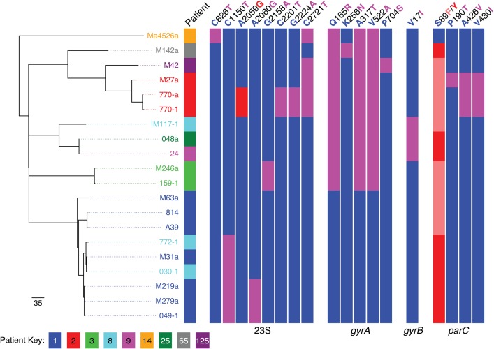
Methods: The bacterial load was estimated in sequential sputum samples from 9 patients by quantitative polymerase chain reaction. The genomes of all available isolates, originating from patients in the United Kingdom, France, and Tunisia, were sequenced along with the type strain. Genomic data were assembled and annotated, and a high-resolution phylogenetic tree was constructed.
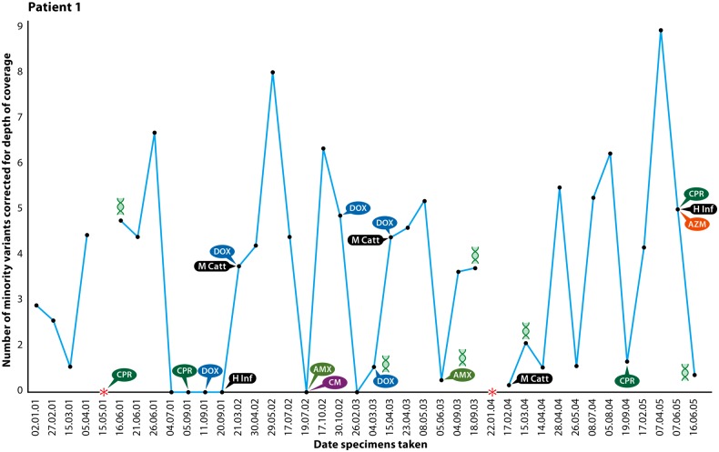
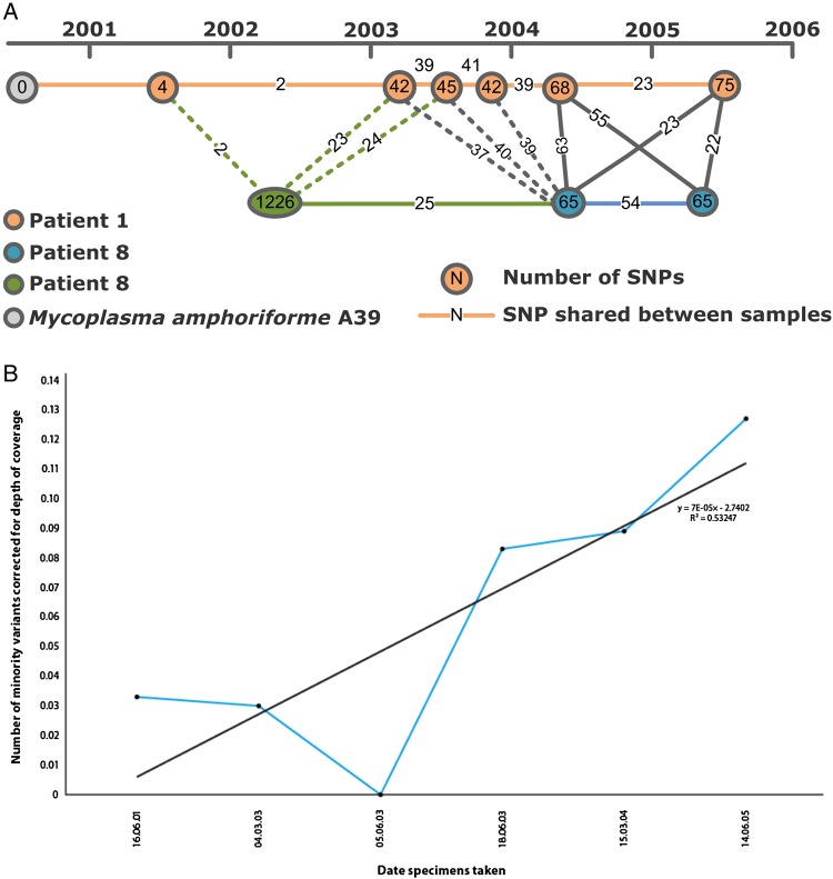
Results: By using high-resolution whole-genome sequencing (WGS) data, we show that patients can be chronically infected with M. amphoriforme manifesting as a relapsing-remitting bacterial load, interspersed by periods when the organism is undetectable. Importantly, we demonstrate transmission of strains within a clinical environment. Antibiotic resistance mutations accumulate in isolates taken from patients who received multiple courses of antibiotics.
Conclusions: Mycoplasma amphoriforme isolates form a closely related species responsible for a chronic relapsing and remitting infection in PAD patients in the United Kingdom and from immunocompetent patients in other countries. We provide strong evidence of transmission between patients attending the same clinic, suggesting that screening and isolation may be necessary for susceptible patients. This work demonstrates the critical role that WGS can play in rapidly unraveling the biology of a novel pathogen.
Keywords: Mycoplasma amphoriforme; infection control; primary antibody deficiency; respiratory infection; whole genome sequencing.
© The Author 2014. Published by Oxford University Press on behalf of the Infectious Diseases Society of America.
Figures
References
-
- Pitcher DG. Mycoplasma amphoriforme sp. nov., isolated from a patient with chronic bronchopneumonia. Int J Syst Evol Microbiol. 2005;55:2589–94. - PubMed
-
- Pereyre S, Renaudin H, Touati A, et al. Detection and susceptibility testing of Mycoplasma amphoriforme isolates from patients with respiratory tract infections. Clin Microbiol Infect. 2009;16:1007–9. - PubMed
-
- Rutherford K, Parkhill J, Crook J, et al. Artemis: sequence visualization and annotation. Bioinformatics. 2000;16:944–5. - PubMed
Publication types
MeSH terms
Grants and funding
LinkOut - more resources
Full Text Sources
Other Literature Sources
Molecular Biology Databases
