. 2014 Dec 5;289(49):34205-13.
doi: 10.1074/jbc.M114.587170. Epub 2014 Oct 24.

E2F transcription factor 1 regulates cellular and organismal senescence by inhibiting Forkhead box O transcription factors

Affiliations

E2F transcription factor 1 regulates cellular and organismal senescence by inhibiting Forkhead box O transcription factors

Qi Xie et al. J Biol Chem. .

Abstract

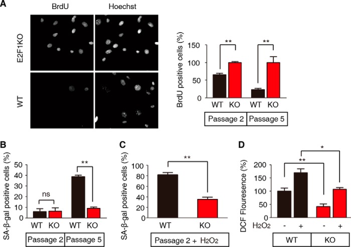
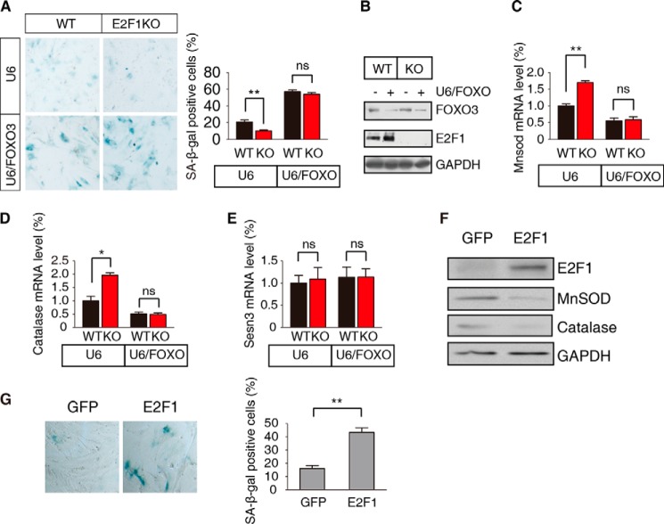
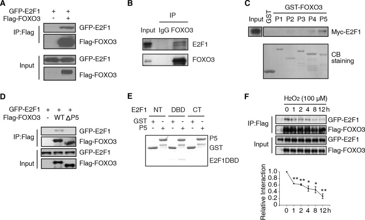
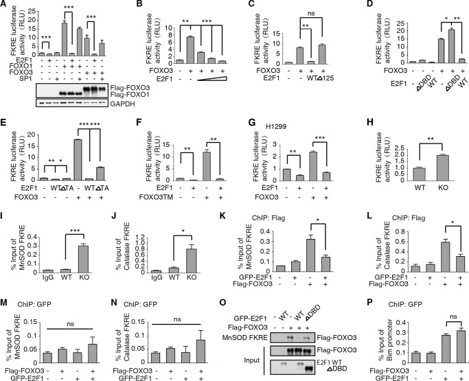
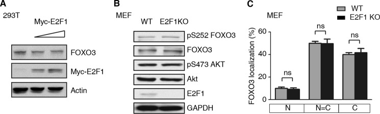
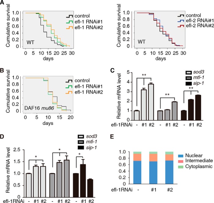
E2F1 and FOXO3 are two transcription factors that have been shown to participate in cellular senescence. Previous report reveals that E2F1 enhanced cellular senescence in human fibroblast cells, while FOXO transcription factors play against senescence by regulation reactive oxygen species scavenging proteins. However, their functional interplay has been unclear. Here we use E2F1 knock-out murine Embryonic fibroblasts (MEFs), knockdown RNAi constructs, and ectopic expression of E2F1 to show that it functions by negatively regulating FOXO3. E2F1 attenuates FOXO3-mediated expression of MnSOD and Catalase without affecting FOXO3 protein stability, subcellular localization, or phosphorylation by Akt. We mapped the interaction between E2F1 and FOXO3 to a region including the DNA binding domain of E2F1 and the C-terminal transcription-activation domain of FOXO3. We propose that E2F1 inhibits FOXO3-dependent transcription by directly binding FOXO3 in the nucleus and preventing activation of its target genes. Moreover, knockdown of the Caenorhabditis elegans E2F1 ortholog efl-1 significantly extends lifespan in a manner that requires the activity of the C. elegans FOXO gene daf-16. We conclude that there is an evolutionarily conserved signaling connection between E2F1 and FOXO3, which regulates cellular senescence and aging by regulating the activity of FOXO3. We speculate that drugs and/or therapies that inhibit this physical interaction might be good candidates for reducing cellular senescence and increasing longevity.

Keywords: E2F Transcription Factor; FOXO; Oxidative Stress; Senescence; Transcription Regulation.


Figures

FIGURE 1.
FIGURE 1.
E2F1 deficiency attenuates MEF senescence. A, proliferation rate of WT and E2F1 KO MEFs at passage 2 and passage 5 as measured by BrdU labeling for 24 h prior to fixation and staining. The left panel shows a representative picture of passage 5 cells visualized by the DNA stain Hoechst and dividing cells by BrdU incorporation. The right panel shows the percent of BrdU-positive cells with the bars representing the mean ± S.E. (t test, **, p < 0.01, n = 3). E2F1 KO cells have been set to 100% to show the percent reduction of division of WT cells. B, WT and E2F1 KO MEFs were stained for senescence-associated β-galactosidase (SA-β-Gal) activity at passage 2 and passage 5. The percentage of SA-β-gal-positive cells was calculated from five randomly chosen fields. At least 150 cells were analyzed per experiment. Data represent the mean ± S.E. (t test, **, p < 0.01, n = 3). C, WT passage 2 MEFs were more sensitive to hydrogen peroxide treatment than E2F1 KO MEFs. Cells were stained for SA-β-Gal activity 48 h after 100 μm H2O2 treatment. Data represent the mean ± S.E. (t test, **, p < 0.01; n = 3). D, ROS levels in WT and E2F1 KO MEFs, treated with hydrogen peroxide or not, were measured by staining with dichlorofluorescein (DCF). Non-treated WT cells have been set to 100%. ROS levels of WT cells are higher than E2F1 KO cells (t test, *, p < 0.05; **, p < 0.01, n = 3). Data represent the mean ± S.E.
FIGURE 2.
FIGURE 2.
E2F1 regulation of senescence requires FOXO3. A, WT and E2F1 KO passage 3 MEFs were infected with lentivirus constitutively expressing FOXO3 (U6/FOXO) shRNA or a control (U6) vector. Senescent cells were examined by SA-β-gal staining and the percent positive cells determined, shown as the mean ± S.E. (t test, **, p < 0.01; ns, not significant, n = 3). B, WT and E2F1 KO MEFs were infected with U6/FOXO or U6 empty lentivirus. Proteins were extracted and detected by immunoblotting with the anti-FOXO3 and E2F1 antibodies. GAPDH serves as the loading control. C-E, WT and E2F1 KO passage 2 MEFs were infected as in A. The mRNA levels of MnSOD (C), catalase (D), and Sesn3 (E) were measured compared relative to WT MEFs infected with control vector. Data are shown as the mean ± S.E. (t test, **, p < 0.01; *, p < 0.05; ns, not significant, n = 3). F, WT passage 3 MEFs were transfected with constitutively expressing GFP-E2F1 (E2F1) or control (GFP) vector. Proteins were extracted and detected by immunoblotting with the indicated antibodies. G, WT passage 3 MEFs were transfected as in F, and senescent cells were visualized by SA-β-gal staining. A representative example is shown in the left panel, and the percent positive cells shown in the right panel as the mean ± S.E. (t test, **, p < 0.01; n = 3).
FIGURE 3.
FIGURE 3.
E2F1 and FOXO3 physically interact in vivo. A, immunoprecipitation of FOXO3 precipitates E2F1. Lysates of HEK293T cells transfected with plasmids encoding GFP-E2F1 and Flag-FOXO3 or control vector were immunoprecipitated with anti-Flag antibody. Western blot were followed with anti-GFP or -Flag antibody. B, immunoprecipitation of endogenous FOXO3 from primary MEFs precipitates E2F1. Anti-FOXO3 or IgG immunoprecipitates from primary MEFs were immunoblotted with FOXO3 or E2F1 antibody. Two bands of endogenous E2F1 were recognized by E2F1 antibody (E2F1 C-20, Santa Cruz Biotechnology). C, The P5 fragment of FOXO3 binds to E2F1 in vitro. GST was fused to 5 different fragments of FOXO3 (P1-P5) and used in a pull-down assay of Myc-E2F1. Precipitated proteins were blotted and detected with the Myc antibody in the upper panel. The lower panel is Coomassie Blue (CB) staining of GST-FOXO3 peptides. D, P5 fragment of FOXO3 is required to bind E2F1. Lysates of HEK293T cells transfected with plasmids encoding GFP-E2F1 and control vector (−), full-length Flag-FOXO3 (WT) or Flag-FOXO3ΔP5 were immunoprecipitated with anti-Flag antibody. Western blot of the immunoprecipitated (top panel) or input (bottom panel) protein were detected with anti-GFP or -Flag antibody. E, P5 fragment of FOXO3 binds a region of E2F1 including its DNA binding domain. GST pull-down assay of His-fusion E2F1 fragments (NT, DBD, CT) with recombinant GST or GST fused with the fragment of FOXO3-P5. F, upper panel: HEK293T was transfected as in (A). 24 h after transfection, 100 μm H2O2 was added into medium. Co-IP assay was performed as in A to show the interaction between FOXO3 and E2F1. Bottom panel: statistical results from three independent experiments. Relative interaction was determined by quantification of GFP-E2F1/Flag-FOXO3 (t test, *, p < 0.05; **, p < 0.01, n = 3).
FIGURE 4.
FIGURE 4.
E2F1 inhibits FOXO3 activity. A, upper panel: FOXO3 and FOXO1 activity is repressed by E2F1. HEK293T cells were transfected with the FOXO3-responsive element, FKRE-luciferase reporter gene, the constitutively active tk-Renilla reporter as a control, and plasmids encoding E2F1 and/or FOXO3/FOXO1,SP1. Data are shown as the mean ± S.E. firefly/Renilla luciferase activity (t test, ***, p < 0.001; n = 3). Bottom Panel: FOXO1 and FOXO3 protein levels were detected by anti-Flag antibody. B, FOXO3 activity is measured as in A. E2F1 expression significantly repressed FOXO3 activity in a dose dependent manner (dose from 0.02–0.1 μg). Data are shown as the mean ± S.E. firefly/Renilla luciferase activity (t test, **, p < 0.01; ***, p < 0.001; n = 3). C, constitutively cytoplasmic E2F1 cannot repress FOXO3 activity. Deletion of the nuclear localization signal in the N-terminal 125 amino acids of E2F1 abolishes the inhibition of FOXO3 by E2F1. HEK293T cells were transfected with FKRE-luciferase and tk-Renilla reporter genes as described in A, together with plasmids encoding full-length (WT) or truncated (Δ125) E2F1 and the FOXO3 expressing plasmid. The data are shown as the mean ± S.E. firefly/Renilla luciferase activity (t test, **, p < 0.01; ns, not significant, n = 3). D, deletion of the FOXO3-binding domain of E2F1 (DBD) abrogates repression. HEK293T cells were transfected as in A with the indicated plasmids. The data are shown as the mean ± S.E. firefly/Renilla luciferase activity (t test, *, p < 0.05; **, p < 0.01; n = 3). E, deletion of the transcription activity domain of E2F1 (E2F1 ΔTA) significantly represses FOXO3a activity. HEK293T cells were transfected as in A with the indicated plasmids. The data are shown as the mean ± S.E. firefly/Renilla luciferase activity (t test, *, p < 0.05; **, p < 0.01; ***, p < 0.001; n = 3). F, E2F1 repression of FOXO3 is independent of Akt phosphorylation. HEK293T cells were transfected as in A except that the Akt independent, activated form of FOXO3, FOXOTM, was used. Data are shown as the mean ± S.E. firefly/Renilla luciferase activity (t test, **, p < 0.01; n = 3). G, E2F1 repression of FOXO3 is independent of p53. The p53 defective cell line H1299 was transfected as in A with indicated plasmids and relative luciferase activity was measured. Data are shown as the mean ± S.E. firefly/Renilla luciferase activity (t test, **, p < 0.01; ***, p < 0.001; n = 3). H, E2F1 KO MEFs have elevated FOXO3 activity. WT and E2F1KO MEFs are transfected with the FKRE-luciferase reporter gene and the tk-Renilla control. Data are shown as the mean ± S.E. firefly/Renilla luciferase activity (t test, **, p < 0.01; n = 3). I–J, FOXO3 enrichment on MnSOD or catalase promoter is elevated in E2F1 KO MEFs. WT and E2F1KO MEFs at passage 5 were cross-linked and subjected to ChIP assay with anti-FOXO3 antibody. Primers corresponding to FKRE of MnSOD or catalase promoter are used to detecting FOXO3 binding. FOXO3 occupancy on MnSOD or catalase increased in E2F1KO MEFs (t test, ***, p < 0.001 on MnSOD promoter, *, p < 0.05 on catalase promoter; n = 3). K–L, FOXO3 enrichment on MnSOD or Catalase promoter is attenuated when E2F1 ectopically expression in HEK293T cells. HEK293T cells were transient transfected with Flag-FOXO3a and/or GFP-E2F1 plasmids as indicated. 24h after transfection, cells were fixed and subjected to ChIP assay with anti-Flag antibody. Primers corresponding to human FKRE of MnSOD or catalase promoter are used to detecting FOXO3 binding. FOXO3a occupancy was decreased on MnSOD and catalase promoter when GFP-E2F1 overexpressed (t test, *, p < 0.05 on MnSOD and catalase promoter, n = 3). M–N, E2F1 enrichment on MnSOD or catalase promoter is not affected by FOXO3a. Cells were transfected as in K and subjected to ChIP assay with anti-GFP antibody. E2F1 binding on FKRE of MnSOD or Catalase promoter were detected qRT-PCR. No significant differences were detected among groups (t test, ns: p > 0.05 on MnSOD and Catalase promoter, n = 3). O, HEK293T cells were transfected with FOXO3 together with GFP-E2F1 WT or ΔDBD. Whole cell extracts were pulled down by biotin-labeled MnSOD FKRE DNA. GFP-E2F1 blocked FOXO3-DNA binding. E2F1 ΔDBD had no significant effect on FOXO3-DNA binding. P, ChIPed DNA from Fig. 4K was analyzed with primers corresponding to E2F1 binding site of Bim promoter. FOXO3 did not affect E2F1 occupancy on Bim promoter.
FIGURE 5.
FIGURE 5.
E2F1 inhibits FOXO activation independent of Akt-mediated phosphorylation and without changing its subcellular localization. A, increasing levels of E2F1 do not affect FOXO3 protein levels. Lysates of HEK293T cells transfected with a constant level of a FOXO3-expressing plasmid and increasing doses Myc-E2F1 were immunoblotted with the indicated antibodies. FOXO3 levels were unchanged relative to an actin control. B, WT and E2F1 KO MEFs have comparable levels of the indicated proteins. Lysates were immunoblotted with the indicated antibodies. C, subcellular localization of GFP-FOXO3 transfected WT or E2F1 KO MEFs is similar. The data are shown as the mean ± S.E. (t test, ns, not significant; n = 3).
FIGURE 6.
FIGURE 6.
E2F (EFL-1) regulation of C. elegans longevity requires FOXO (DAF-16). A and B, reducing expression of EFL-1 by feeding wild-type worms bacteria expressing either of two different RNAi efl-1 clones, compared with a control clone, extended lifespan (A, left), (**, p < 0.001 for efl-1#1 or efl-1#2 RNAi versus control RNAi), but not efl-2 RNAi (A, right) (not significant for efl-2#1 or efl-2#2 RNAi versus control RNAi). Daf-16 (mu86), a putative null mutation of FOXO, doesn't extend lifespan, when knockdown efl-1 (B) (not significant for efl-1#1 or efl-1#2 RNAi versus control RNAi). C–D, increased longevity of efl-1 knockdown worms correlates with increased levels of stress response genes sod3, mtl-1, and sip1. Relative mRNA levels of the indicated genes were determined by quantitative real-time qPCR in wild type (C) (t test, **, p < 0.01; n = 3) and daf-16 (mu86) worms (D) (t test, *, p < 0.05; n = 3), fed bacteria as in A and B. E, GFP-DAF-16 transgenic worms were used for subcellular localization analysis of DAF-16. EFL-1 did not affect the subcellular localization of DAF-16 in worms (not significant for efl-2#1 or efl-2#2 RNAi versus control RNAi).

Similar articles

Cited by

References

    1. Wu Z., Zheng S., Yu Q. (2009) The E2F family and the role of E2F1 in apoptosis. Int. J. Biochem. Cell Biol. 41, 2389–2397 - PubMed
    1. Matsumura I., Tanaka H., Kanakura Y. (2003) E2F1 and c-Myc in cell growth and death. Cell Cycle 2, 333–338 - PubMed
    1. Dimri G. P., Itahana K., Acosta M., Campisi J. (2000) Regulation of a senescence checkpoint response by the E2F1 transcription factor and p14(ARF) tumor suppressor. Mol. Cell. Biol. 20, 273–285 - PMC - PubMed
    1. DeGregori J., Kowalik T., Nevins J. R. (1995) Cellular targets for activation by the E2F1 transcription factor include DNA synthesis- and G1/S-regulatory genes. Mol. Cell. Biol. 15, 4215–4224 - PMC - PubMed
    1. Field S. J., Tsai F. Y., Kuo F., Zubiaga A. M., Kaelin W. G., Jr., Livingston D. M., Orkin S. H., Greenberg M. E. (1996) E2F-1 functions in mice to promote apoptosis and suppress proliferation. Cell 85, 549–561 - PubMed

Publication types

MeSH terms