Glomus tumor of the trachea managed by spiral tracheoplasty
- PMID: 25344687
- PMCID: PMC4214701
- DOI: 10.12659/AJCR.891191
Glomus tumor of the trachea managed by spiral tracheoplasty
Abstract
Background: Glomus tumors are usually found over the dermis of the extremities, particularly over the subungual region of the fingers, and occurrence in the trachea is an extremely rare event. To date, only 29 cases of tracheal and 2 main bronchus glomus tumors have been reported in the English literature. Our patient is the first ever reported case in Taiwan that was managed by spiral tracheoplasty.
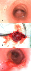
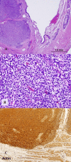
Case report: A 58-year-old woman was admitted to our hospital because of hemoptysis. Computed tomographic (CT) scan revealed a mass over the posterior wall of the trachea. Surgical resection with spiral tracheoplasty was performed due to uncontrolled bleeding and airway compromise. Histopathology and immunostaining confirmed a glomus tumor. Postoperative course was unremarkable and she was discharged in improved condition after 9 days of hospital stay.
Conclusions: Although chronic symptom presentation is the rule for tracheal glomus tumors, airway obstruction and bleeding are life-threatening presentations. Histopathological examination and staining are important to differentiate it from hemangiopericytoma or carcinoid tumors. Spiral tracheoplasty after tangential resection may be tried, as this preserves more tracheal tissue, decreases tension, and prevents postoperative leakage at the anastomotic site.
Figures


Similar articles
-
Glomus Tumor of Trachea in an Adult Male.J Coll Physicians Surg Pak. 2016 Jun;26(6 Suppl):S59-60. J Coll Physicians Surg Pak. 2016. PMID: 27376225
-
Nonintubated Spontaneous Respiration Anesthesia for Tracheal Glomus Tumor.Ann Thorac Surg. 2017 Aug;104(2):e161-e163. doi: 10.1016/j.athoracsur.2017.02.028. Ann Thorac Surg. 2017. PMID: 28734442
-
Glomus tumor of the trachea.J Bronchology Interv Pulmonol. 2012 Jul;19(3):220-3. doi: 10.1097/LBR.0b013e31825ceef8. J Bronchology Interv Pulmonol. 2012. PMID: 23207466
-
Malignant glomus tumor of trachea: a case report with literature review.Asian Cardiovasc Thorac Ann. 2016 Jan;24(1):104-6. doi: 10.1177/0218492315608546. Epub 2015 Sep 28. Asian Cardiovasc Thorac Ann. 2016. PMID: 26420909 Review.
-
Glomus tumor of the trachea.Ann Thorac Surg. 2000 Jul;70(1):295-7. doi: 10.1016/s0003-4975(00)01285-6. Ann Thorac Surg. 2000. PMID: 10921732 Review.
Cited by
-
The mimic of tracheal carcinoid in elderly female.Lung India. 2018 Jan-Feb;35(1):47-49. doi: 10.4103/0970-2113.221726. Lung India. 2018. PMID: 29319034 Free PMC article.
-
Glomus tumors of the trachea: 2 case reports and a review of the literature.J Thorac Dis. 2017 Sep;9(9):E815-E826. doi: 10.21037/jtd.2017.08.54. J Thorac Dis. 2017. PMID: 29221350 Free PMC article.
-
Rare airway tumors: an update on current diagnostic and management strategies.J Thorac Dis. 2016 Aug;8(8):1922-34. doi: 10.21037/jtd.2016.07.40. J Thorac Dis. 2016. PMID: 27621844 Free PMC article. No abstract available.
-
Successful Resection of locally infiltrative Glomus Tumor without pulmonary resection.Int J Surg Case Rep. 2017;41:191-193. doi: 10.1016/j.ijscr.2017.09.015. Epub 2017 Sep 23. Int J Surg Case Rep. 2017. PMID: 29096341 Free PMC article.
-
Glomus tumor of the trachea: a rare case report.Int J Clin Exp Pathol. 2015 Aug 1;8(8):9723-6. eCollection 2015. Int J Clin Exp Pathol. 2015. PMID: 26464745 Free PMC article.
References
-
- Hussarek M, Reider W. Glomus tumor the air tubes. Krebsarzt. 1950;5:208–12. - PubMed
-
- Fabich DR, Hafez GR. Glomangioma of the trachea. Cancer. 1980;45:2337–41. - PubMed
-
- Warter A, Vetter JM, Morand G, Philippe E. Tracheal glomus tumor. Arch Anat Cytol Pathol. 1980;28:184–90. - PubMed
-
- Ito H, Motohiro K, Nomura S, Tahara E. Glomus tumor of the trachea: immunohistochemical and electron microscopic studies. Pathol Res Pract. 1988;183:778–84. - PubMed
Publication types
MeSH terms
LinkOut - more resources
Full Text Sources
Other Literature Sources
Medical

