. 2014 Dec;16(12):1227-37.
doi: 10.1038/ncb3054. Epub 2014 Oct 26.

Rsp5/Nedd4 is the main ubiquitin ligase that targets cytosolic misfolded proteins following heat stress

Affiliations

Rsp5/Nedd4 is the main ubiquitin ligase that targets cytosolic misfolded proteins following heat stress

Nancy N Fang et al. Nat Cell Biol. 2014 Dec.

Abstract

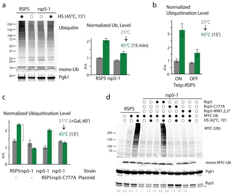
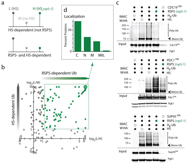
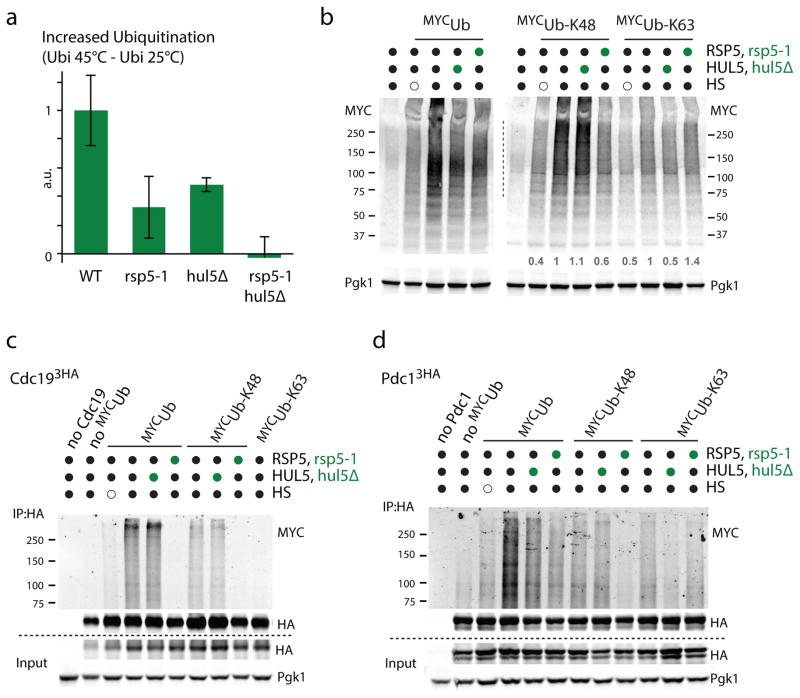
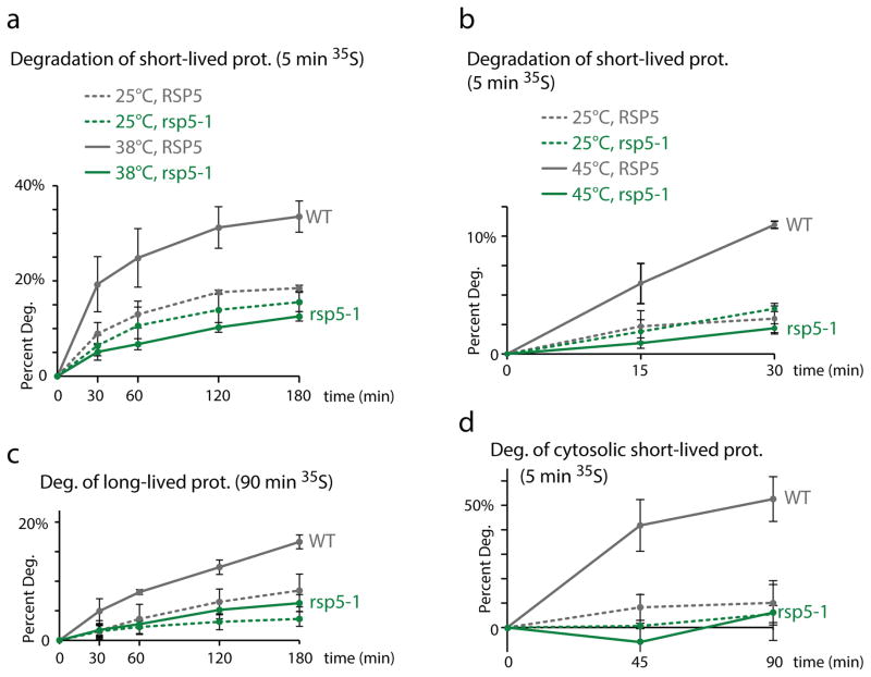
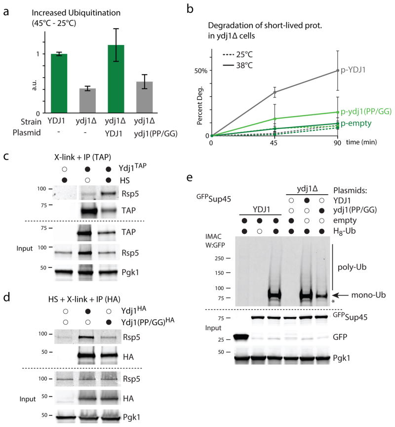
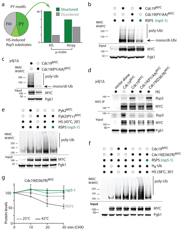
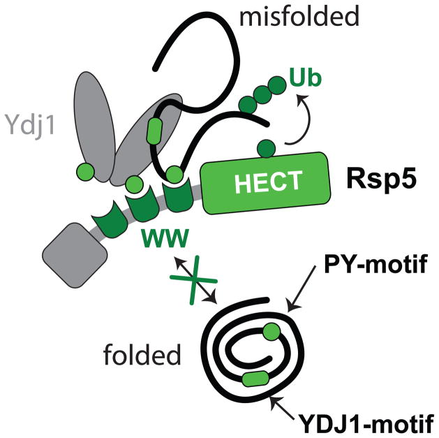
The heat-shock response is a complex cellular program that induces major changes in protein translation, folding and degradation to alleviate toxicity caused by protein misfolding. Although heat shock has been widely used to study proteostasis, it remained unclear how misfolded proteins are targeted for proteolysis in these conditions. We found that Rsp5 and its mammalian homologue Nedd4 are important E3 ligases responsible for the increased ubiquitylation induced by heat stress. We determined that Rsp5 ubiquitylates mainly cytosolic misfolded proteins upon heat shock for proteasome degradation. We found that ubiquitylation of heat-induced substrates requires the Hsp40 co-chaperone Ydj1 that is further associated with Rsp5 upon heat shock. In addition, ubiquitylation is also promoted by PY Rsp5-binding motifs found primarily in the structured regions of stress-induced substrates, which can act as heat-induced degrons. Our results support a bipartite recognition mechanism combining direct and chaperone-dependent ubiquitylation of misfolded cytosolic proteins by Rsp5.


Conflict of interest statement

The authors declare no competing financial interests.

Figures

Figure 1
Figure 1. Rsp5 is a major E3 that mediates the increased ubiquitination upon HS
a. Ubiquitination levels in both WT and rsp5-1 cells before (empty circle) and after a direct HS (black) analyzed by western blots and quantified by dot blot (right; see also Supplementary Figure 1a). Free mono-ubiquitin detected on the lower portion of the gel and Pgk1 loading-control are also shown. b. Normalized ubiquitination levels quantified by dot-blot in Tetp::RSP5 cells before and after HS (see also Supplementary Figure 1b). c. Normalized ubiquitination levels before and after HS in WT and rsp5-1 cells containing the indicated plasmids (- denotes presence of a control empty plasmid). Both RSP5 and rsp5-C777A were expressed under a Gal-promoter induced for 60min with 2% Galactose at 37°C. In a-c, a.u. denotes arbitrary units (each value is relative to the averaged value of the reference sample), n=3 (average±SD; source values are listed in Supplementary Table 5). d. In vitro ubiquitination performed with the indicated (black circles) recombinant proteins in WT and rsp5-1 cell extracts incubated at 25°C (empty circle) or 42°C for 10min (HS; black) and analyzed by western blot. Mono- MYC-ubiquitin, Rsp5 (with relative quantified revels) and Pgk1 loading control are also shown. The asterisk denotes an unspecific band. All uncropped images are in Supplementary Figure 8.
Figure 2
Figure 2. The role of Rsp5/Nedd4 in the increased ubiquitination upon HS is conserved
a. Ubiquitination levels in the indicated heat-stressed and unstressed HeLa cells stably knocked-down for Nedd4 (clones #A and #B), or scrambled control (−), as represented by the immunoblot in the left panel. Quantification of three experiments is provided in the right panel (average±SD; source values are listed in Supplementary Table 5). b. As in a, only Nedd4 knockout (−/−) or WT (+/+) MEFs were used instead of HeLa cells. c. As in a, but with the stably knocked-down HeLa cells re-transfected (or mock transfected) with WT NEDD4 or the catalytically-inactive NEDD4(C867S) for 24hrs prior to HS (45°C, 30min). All uncropped images are in Supplementary Figure 8.
Figure 3
Figure 3. Rsp5 targets mainly cytosolic proteins upon HS
a. Schematic representation of the triple SILAC experiment to identify HS-induced Rsp5 substrates (L:light; M:medium; and H: high labels). b. Plot of the log2 ratios of each quantified ubiquitinated peptide. In the y-axis, ratios of L versus M are reported to display sites affected by HS, and ratios of L versus H are reported in the x-axis to indicate sites affected by the absence of Rsp5 activity. RSP5-dependent ubiquitination sites are indicated in green. The are 76 overlapping sites with log2(ratios) ≥ 5.64. c. IMAC-enriched ubiquitin conjugates were analyzed by western blots. Samples were derived from indicated cells (rsp5-1 designated by green circles) was assed expressing endogenously tagged candidates (3xHA) and plasmid version of H8-Ubiquitin. All uncropped images are in Supplementary Figure 8. d. Localization of HS-induced Rsp5 substrates identified in b (C: Cytoplasm; N: Nuclear; M: Membrane; Mit: Mitochondria; source values are listed in Supplementary Table 5).
Figure 4
Figure 4. Both Rsp5 and Hul5 mostly target their substrates independently
a. Increased ubiquitination levels quantified by dot-blot after a 15min HS at 45°C in the indicated cells. Experiment was done with three biological replicates (average±SD; source values are listed in Supplementary Table 5) and a.u. denotes arbitrary units. b. Western blot analysis of the indicated cells that expressed on a plasmid the indicated N-terminally MYC tagged ubiquitin constructs (K48 and K63 designate ubiquitin variants that only contain K48 and K63, respectively, while all other lysines are mutated to arginines) that were subjected or not to HS (45°C, 15min). c–d. C-terminally 3xHA tagged Cdc19 (c) and Pdc1 (d) expressed from their endogenous promoter on a plasmid were immunoprecipitated from the indicated cells (hul5Δ and rsp5-1 designated by green circles) expressing the indicated N-terminally MYC tagged ubiquitin constructs. Cells were HS or not for 20min at 45°C. Wild-type MYC tagged ubiquitin (MYCUb) was also expressed in the control cells used in lane 1. All uncropped images are in Supplementary Figure 8.
Figure 5
Figure 5. Rsp5 is required for the degradation of cytosolic misfolded proteins
a–d. Degradation of 35S labeled proteins in WT (grey) and rsp5-1 (green) cells at 25°C (dotted lines) or upon HS (solid lines; 38°C in a, c and d and 45°C in b). The portion of proteins degraded after the chase at the indicated times was measured for short-lived (a, b) and long-lived (c) proteins. The portion of proteins that mainly correspond to cytosolic proteins degraded after the chase was measured at the indicated times in (d). All experiments were done in three independent experiments (average±SD; source values are listed in Supplementary Table 5).
Figure 6
Figure 6. An Ydj1-adaptor mediates ubiquitination of misfolded proteins upon HS
a. Increased ubiquitination levels quantified by dot-blot after HS (45°C, 15min) in the indicated cells that carried a LEU2 plasmid that was empty (−) or with YDJ1 or ydj1 (PP/GG) expressed from the YDJ1 promoter. Three biological replicates were assessed (average±SD; source values are listed in Supplementary Table 5). b. Degradation of short-lived 35S-labeled (5 min) proteins in ydj1Δ cells that carried a plasmid expressing YDJ1 (grey) or the ydj1 (PP/GG) mutant (light green) from the YDJ1 promoter or an empty plasmid (dark green) at 25°C (dotted lines) or 38°C (solid lines). Three independent experiments were performed (average±SD; source values are listed in Supplementary Table 5). c. TAP immunoprecipitation (IP) was performed after in vivo cross-linking with 1% formaldehyde in cells grown at 25°C (empty circle) or during a HS (45°C, 20min; black). The endogenous YDJ1 was C-terminally TAP-tagged in the indicated lanes. The IP samples were analyzed by western blot (the - TAP control was analyzed on the same membrane but not adjacent to the other two lanes). d. HA IP was performed after in vivo cross-linking with 1% formaldehyde in ydj1Δ cells subjected to a HS (45°C, 20min) and that carried a LEU2 plasmid that was empty or contained C-terminally tagged (3xHA) YDJ1 or ydj1 (PP/GG) expressed from the YDJ1 promoter. e. IMAC from YDJ1 or ydj1Δ cells expressing when indicated H8-Ubiquitin from a first URA3 plasmid, Sup45 fused N-terminally to GFP or GFP alone from a second HIS3 plasmid, and YDJ1 or ydj1 (PP/GG) under the YDJ1 promoter from third LEU2 plasmid. All uncropped images are in Supplementary Figure 8.
Figure 7
Figure 7. PY-motifs on substrates promote RSP5-dependent ubiquitination upon HS
a. The pie chart (left) indicates which portion of HS induced Rsp5 candidate substrates identified in the two mass spectrometry experiments contains a proline-containing motifs ([PLSV]Px[YF], or PPPP; dark green), additional PxY motifs (light green) or no obvious PY motif (grey). × denotes any residue. The histogram (right) indicates whether [PLSV]Px[YF] or PPPP motifs are located in regions predicted disordered or not among Rsp5 candidate substrates induced by HS or identified in a protein array (Array). Assigned PY motifs are listed in Supplementary Table 5. b, c, e, f. IMAC analyzed by western blots. Samples were derived from cells expressing H8-ubiquitin (from a URA3 plasmid) and the indicated wild-type or mutants Cdc19 and Pyk2 (fused C-terminally with 13xMYC and expressed from a HIS3 plasmid). Py+ designates the A365P/L366N mutations in Pyk2. RSP5 (black) or rsp5-1 (green) cells were analyzed in b, e and f, and ydj1Δ cells in c. A HS at 45°C for 20min was applied in b and c, and in e when indicated (black); cells were subjected to a 38°C HS for 30min in f when indicated (black). d. MYC immunoprecipitation (IP) was performed after in vivo cross-linking with 1% formaldehyde (10 min) in cells grown at 25°C (empty circle) or during a HS (40°C, 20min; black). The indicated Cdc19 (fused C-terminally with 13xMYC) were expressed (GDP promoter) in ydj1Δ cells. The IP and input samples were analyzed by western blots as indicated. All uncropped images are in Supplementary Figure 8. g. Levels of Cdc19(D367R) C-terminally tagged with 13×MYC was determined by quantitative western blotting after incubating cells (wild type in grey and rsp5-1 in green) in the presence of 100μg/ml cycloheximide at 25°C (dotted lines) or 42°C (solid lines) at the indicated times in three independent experiments (average±SEM; source values are listed in Supplementary Table 5; representative images are shown in Supplementary Figure 7f).
Figure 8
Figure 8. Bipartite model for the recognition of cytosolic misfolded proteins by Rsp5
Schematic of the proposed bipartite model in which misfolded substrates are recruited by Ydj1 Hsp40 and/or directly recognized by Rsp5.

Comment in

Similar articles

Cited by

References

    1. Kim YE, Hipp MS, Bracher A, Hayer-Hartl M, Hartl FU. Molecular chaperone functions in protein folding and proteostasis. Annu Rev Biochem. 2013;82:323–355. - PubMed
    1. Powers ET, Balch WE. Diversity in the origins of proteostasis networks--a driver for protein function in evolution. Nat Rev Mol Cell Biol. 2013;14:237–248. - PMC - PubMed
    1. Chen B, Retzlaff M, Roos T, Frydman J. Cellular strategies of protein quality control. Cold Spring Harb Perspect Biol. 2011;3:a004374. - PMC - PubMed
    1. Comyn SA, Chan GT, Mayor T. False start: Cotranslational protein ubiquitination and cytosolic protein quality control. J Proteomics. 2013 - PubMed
    1. Brodsky JL. Cleaning up: ER-associated degradation to the rescue. Cell. 2012;151:1163–1167. - PMC - PubMed

Publication types

MeSH terms

Substances