Comparative Study
. 2014 Dec;74(6):1297-305.
doi: 10.1007/s00280-014-2610-x. Epub 2014 Oct 26.

Addition of bevacizumab enhances antitumor activity of erlotinib against non-small cell lung cancer xenografts depending on VEGF expression

Affiliations
Comparative Study

Addition of bevacizumab enhances antitumor activity of erlotinib against non-small cell lung cancer xenografts depending on VEGF expression

Heyan Li et al. Cancer Chemother Pharmacol. 2014 Dec.

Abstract

Purpose: Erlotinib, an epidermal growth factor receptor (EGFR) tyrosine kinase inhibitor (TKI), and bevacizumab, an anti-vascular endothelial growth factor (VEGF) agent, are promising therapies for advanced non-small cell lung cancer (NSCLC). Our study was aimed to determine whether there were conditions under which the addition of bevacizumab would enhance the antitumor activity of erlotinib against NSCLC tumors in vitro and in vivo.

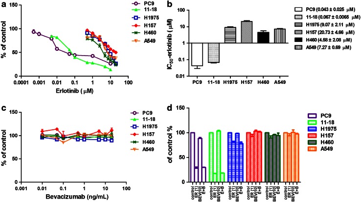
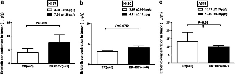
Methods: MTS was for NSCLC cell (PC9, 11-18, H1975, H157, H460 and A549) growth assay in vitro. ELISA was for VEGF protein assay in cells and tumor tissues. Mouse xenograft models were established with H157, H460 and A549 with primary resistance to erlotinib and treated with erlotinib plus bevacizumab or each agent alone. Erlotinib concentrations in tumors were determined by high-performance liquid chromatography.

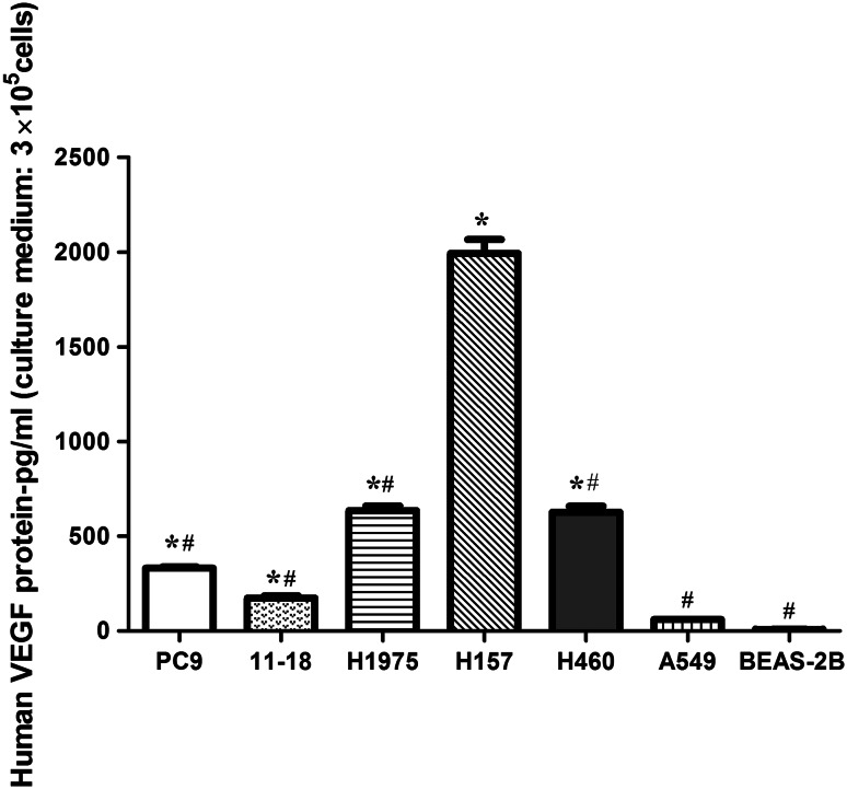
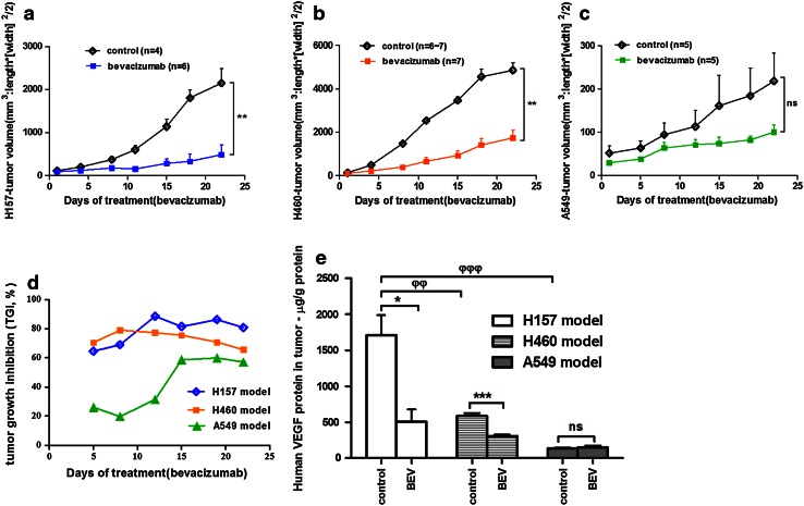
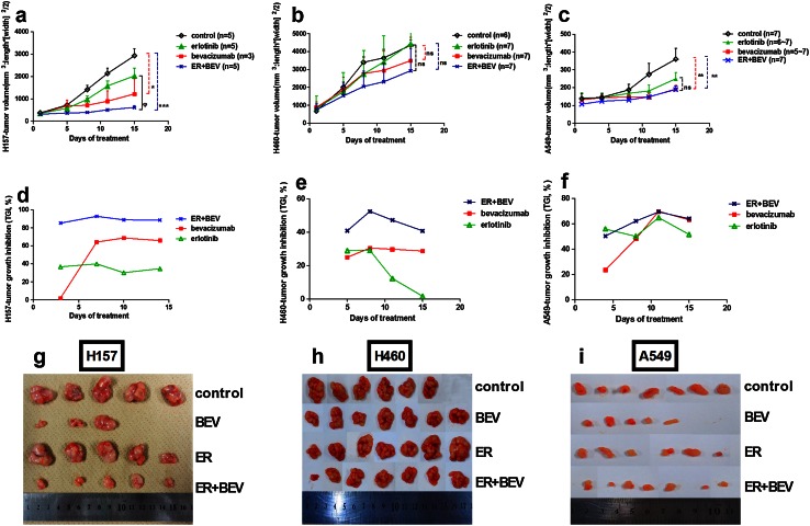
Results: Bevacizumab alone did not inhibit NSCLC cell growth in vitro. In primarily erlotinib-resistant NSCLC cells, the levels of VEGF protein were highest in H157 cell followed in order by H460 and A549 cells. In vivo, bevacizumab alone significantly inhibited tumor growth only in xenograft models with high (H157) and/or moderate (H460) levels of VEGF protein. A combination of erlotinib and bevacizumab partially reversed resistance to erlotinib in H157 xenografts (high VEGF level) with increasing intratumoral erlotinib concentrations, but not in H460 (moderate) or A549 (low) xenografts.

Conclusions: These results support that combined with anti-VEGF therapy could enhance antitumor activity of anti-EGFR therapy and/or partially reverse resistance to EGFR TKI, by increasing EGFR TKI concentration in specific tumors that express high levels of VEGF protein.


Figures

Fig. 1
Fig. 1
Effects of erlotinib/bevacizumab on NSCLC cell lines in vitro. MTS assay was used to evaluate the effects of erlotinib (a), bevacizumab (c), and combination of erlotinib and bevacizumab (d) on the growth of NSCLC cell lines, which included cell lines with EGFR mutations: PC9 (EGFR exon 19 deletion), 11–18 (EGFR L858), H1975 (EGFR L858R and T790 M mutations) and EGFR wild-type cell lines: H157, H460 and A549. Cells were treated with erlotinib (0–20 µmol/L), bevacizumab (0–20 ng/mL) or combination of these agents (ER 1 µmol/L; BEV 10 ng/mL) for 72 h. The percentage of viable cells is shown relative to that of the untreated control. b The IC50 of erlotinib in the different cell lines. d No significant differences were noted between erlotinib alone and combination treatment in vitro (P > 0.05). Results are presented as the mean ± SEM. ER erlotinib, BEV bevacizumab
Fig. 2
Fig. 2
Levels of human VEGF protein in NSCLC cell lines. Human VEGF protein in culture medium (2 mL with free fetal bovine serum) of NSCLC cell lines (3 × 105 cells) and the human bronchial epithelial cell line BEAS-2B (control) was assessed by ELISA. Data are presented as the mean ± SEM. *P < 0.05 for cells compared with BEAS-2B; # P < 0.05 for cells compared with H157
Fig. 3
Fig. 3
Effects of bevacizumab monotherapy on NSCLC xenograft models with primary resistance to erlotinib. a, b and c Tumor volume over time in response to bevacizumab (5 mg/kg; n = 4–7). The Mann–Whitney U test was used to compare tumor volume at the last measurement between the groups (ΔT/ΔC): **P < 0.01 in H157 tumors (a) and H460 tumors (b); ns P > 0.05 in A549 tumors (c). d TGI  % in each model was calculated from the beginning of bevacizumab treatment. e The levels of human VEGF protein in tumor tissues of H157, H460 and A549 models were assayed by ELISA. The Student’s t test was used to compare bevacizumab and vehicle treatment in each model: *P < 0.05, ***P < 0.001, ns P > 0.05. For comparison between the three xenograft tumors, one-way ANOVA was used: φφ P < 0.01, φφφ P < 0.001. Data are expressed as the mean ± SEM. ER erlotinib, BEV bevacizumab
Fig. 4
Fig. 4
Effects of erlotinib plus bevacizumab on tumor growth in NSCLC xenograft models. a, b and c Changes in tumor volume over time in response to treatment with vehicle, bevacizumab (5 mg/kg), erlotinib (100 mg/kg) or combination of bevacizumab (5 mg/kg) and erlotinib (100 mg/kg) for 2 weeks (n = 3–7). One-way ANOVA was used to compare tumor volume at the last measurement between the treatment groups in each xenograft model: H157 model (a), H460 model (b), and A549 model (c). ***P < 0.001, *P < 0.05, **P < 0.01 for combination treatment or bevacizumab treatment compared with the vehicle (ΔT/ΔC); φ P < 0.05 for combination treatment compared with erlotinib alone (ΔT/ΔT′); and ns P > 0.05 means no significant differences. d, e and f TGI  % by erlotinib, bevacizumab and combination treatment in three models. g, h and i Images of tumor samples in three xenografts. Data are expressed as the mean ± SEM. ER erlotinib, BEV bevacizumab
Fig. 5
Fig. 5
Erlotinib concentration in tumor tissues. HPLC was used to determine the erlotinib concentration in the tumor tissues from the xenograft models treated with erlotinib alone or combined with bevacizumab. Data are expressed as the mean ± SEM. The Student’s t test was used to compare erlotinib and combination groups in each model: P = 0.289 (a H157 model), P = 0.0751 (b H460 model) and P = 0.569 (c A549 model). ER erlotinib, BEV bevacizumab

Similar articles

Cited by

References

    1. Gerber DE. Targeted therapies: a new generation of cancer treatments. Am Fam Physician. 2008;77(3):311–319. - PubMed
    1. Jemal A, Bray F, Center MM, Ferlay J, Ward E, Forman D. Global cancer statistics. CA-Cancer J Clin. 2011;61(2):69–90. doi: 10.3322/caac.20107. - DOI - PubMed
    1. Siegel R, Naishadham D, Jemal A. Cancer statistics, 2013. CA-Cancer J Clin. 2013;63(1):11–30. doi: 10.3322/caac.21166. - DOI - PubMed
    1. Custodio A, Mendez M, Provencio M. Targeted therapies for advanced non-small-cell lung cancer: current status and future implications. Cancer Treat Rev. 2012;38(1):36–53. doi: 10.1016/j.ctrv.2011.04.001. - DOI - PubMed
    1. Cohen MH, Johnson JR, Chen YF, Sridhara R, Pazdur R. FDA drug approval summary: erlotinib (Tarceva (R)) tablets. Oncologist. 2005;10(7):461–466. doi: 10.1634/theoncologist.10-7-461. - DOI - PubMed

Publication types

MeSH terms