. 2014 Oct 15;5(19):9007-21.
doi: 10.18632/oncotarget.2346.

A novel calcium-dependent mechanism of acquired resistance to IGF-1 receptor inhibition in prostate cancer cells

Affiliations

A novel calcium-dependent mechanism of acquired resistance to IGF-1 receptor inhibition in prostate cancer cells

Cale D Fahrenholtz et al. Oncotarget. .

Abstract

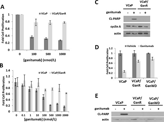
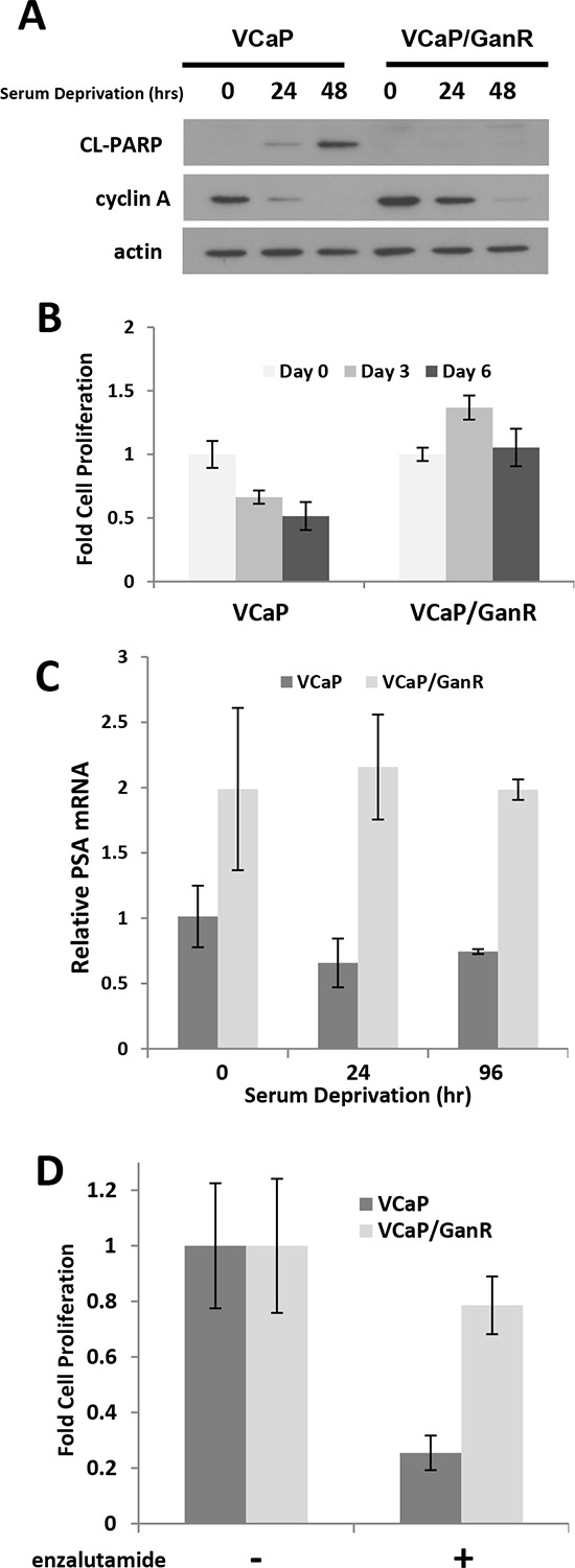
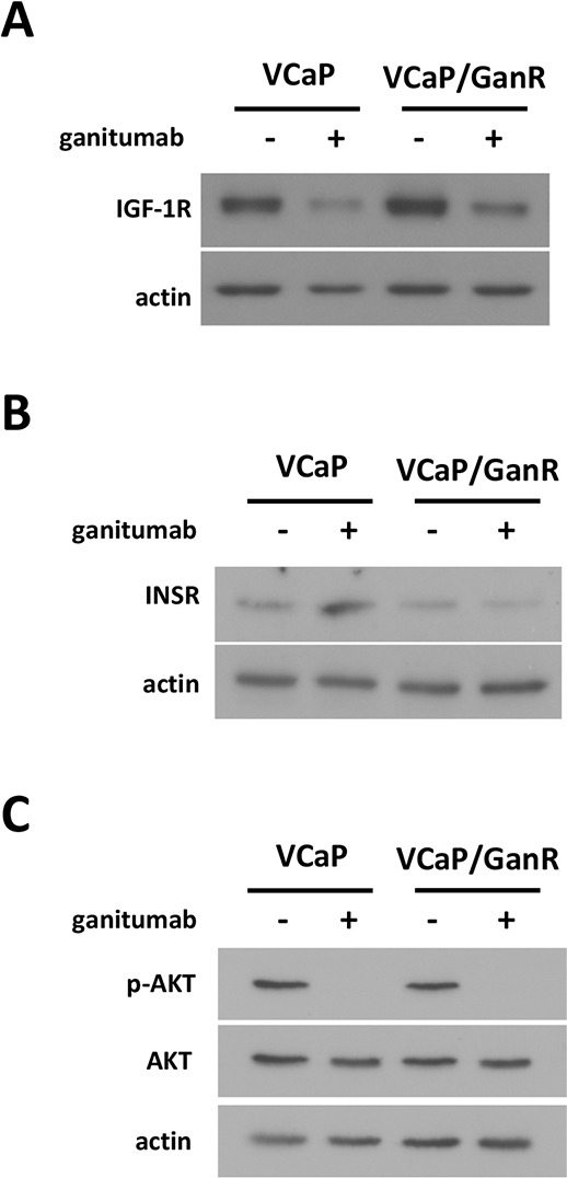
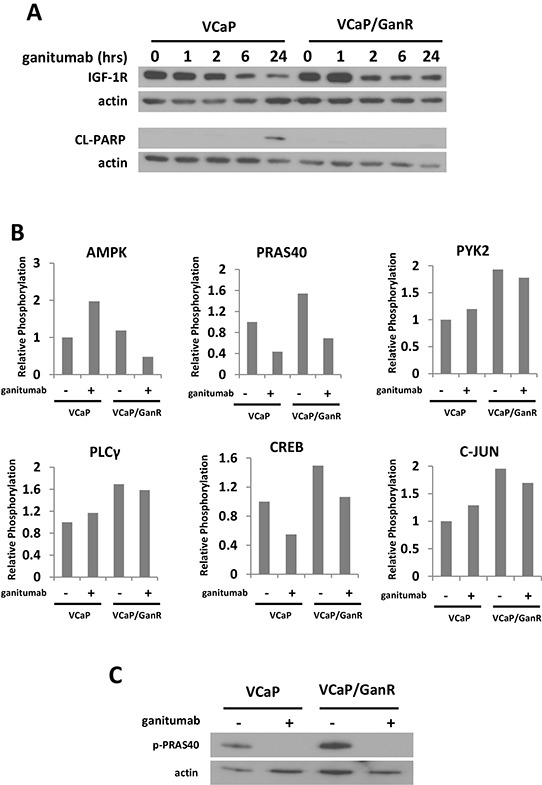
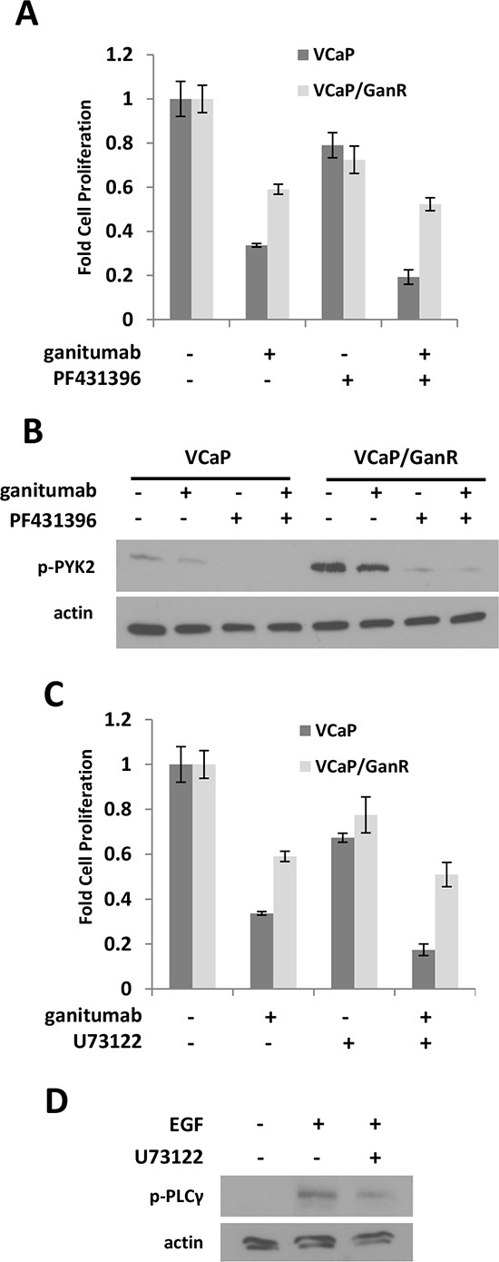
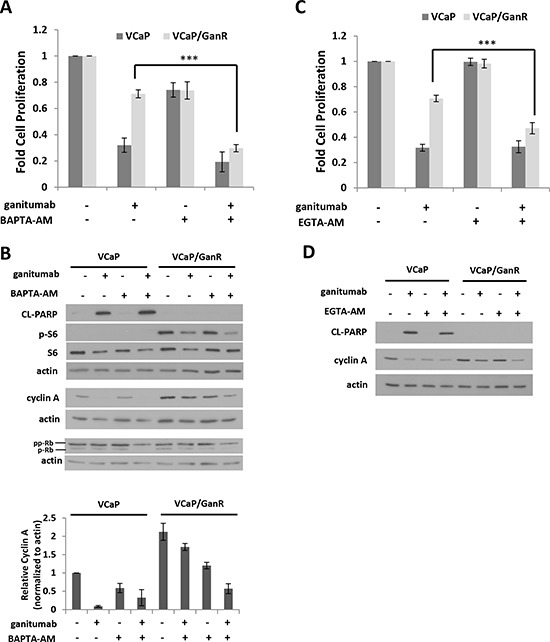
Inhibition of the mitogenic insulin-like growth factor receptor 1 (IGF-1R) signaling axis is a compelling treatment strategy for prostate cancer. Combining the IGF-1R inhibitor ganitumab (formerly AMG 479) with standard of care androgen-deprivation therapy greatly delays prostate cancer recurrence in xenograft models; however, a significant proportion of these tumors ultimately acquire resistance to ganitumab. Here we describe the development of a stable and reproducible ganitumab-resistant VCaP human prostate cancer cell derivative termed VCaP/GanR to investigate the mechanism of acquired resistance to IGF-1R inhibition. Unlike parental VCaP, VCaP/GanR did not undergo apoptosis following ganitumab treatment. VCaP/GanR did not express increased levels of IGF-1R, insulin receptor, or phospho-AKT compared to parental VCaP. VCaP/GanR exhibited increased levels of phospho-S6 indicative of increased mTOR activity. However, acquired resistance to ganitumab was not dependent on increased mTOR activity in VCaP/GanR. Phospho-proteomic arrays revealed alterations in several calcium-regulated signaling components in VCaP/GanR compared to VCaP. Reduction of intracellular calcium using cell-permeable calcium-specific chelators restored ganitumab sensitivity to VCaP/GanR through inhibition of cell-cycle progression. These data suggest a new mechanism of resistance to IGF-1R inhibition involving calcium-mediated proliferation effects. Such pathways should be considered in future clinical studies of IGF-1R inhibitors in prostate cancer.


Conflict of interest statement

Disclosure of Potential Conflicts of Interest

Pedro J. Beltran is an employee of and owns stock in Amgen, Inc.

Figures

Figure 1
Figure 1. Characterization of a ganitumab resistant derivative of human prostate cancer VCaP termed VCaP/GanR
VCaP and VCaP/GanR were treated with ganitumab (A) (0–1000 nmol/L) or (B) (0–2000 nmol/L) for six days in medium containing 2% FBS and proliferation relative to vehicle control is shown. (C) VCaP and VCaP/GanR were treated with ganitumab (500 nmol/L) or vehicle in medium containing 2% FBS for 72 hours and lysates were probed for cleaved PARP, cyclin A and actin. (D) VCaP, VCaP/GanR and VCaP/GanWD were treated with ganitumab (500 nmol/L) or vehicle for six days in medium containing 2% FBS and cell proliferation relative to vehicle treatment is shown. (E) VCaP, VCaP/GanR and VCaP/GanWD were treated with ganitumab (500 nmol/L) or vehicle in medium containing 2% FBS for 72 hours and probed for cleaved PARP and actin. Panel (A) represents three combined independent experiments performed in triplicate. Panels (B-E) are representative of at least 2 independent experiments. Data are shown ± SD.
Figure 2
Figure 2. Castration-resistant characteristics of VCaP/GanR
(A) VCaP and VCaP/GanR were cultured under androgen- and mitogen-depleted conditions in medium containing 10% CSS for the indicated times, and probed for cleaved PARP, cyclin A, and actin. (B) VCaP and VCaP/GanR were cultured in medium supplemented with 10% CSS and cell proliferation is shown relative to cell number at initiation of serum deprivation (Day 0). (C) VCaP and VCaP/GanR were grown in medium containing 10% CSS for the times indicated. PSA mRNA was assessed by reverse transcriptase realtime PCR, normalized to HPRT, and shown relative to parental VCaP. (D) VCaP and VCaP/GanR were treated for five days with androgen receptor inhibitor enzalutamide (500 nmol/L) in medium containing 2% FBS and proliferation is shown relative to vehicle treatment. Panels (B-D) were performed in triplicate. Data are shown ± SD.
Figure 3
Figure 3. Characterization of IGF-1R-related signaling pathways in VCaP/GanR
VCaP and VCaP/GanR were treated with ganitumab (500 nmol/L) or vehicle for 72 hours in medium containing 2% FBS. Lysates were immunoblotted for IGF-1R and actin (A), INSR and actin (B), or phospho-AKT then probed for total AKT and actin (C). Panels (A,B,C) are representative of three independent experiments.
Figure 4
Figure 4. Phospho-proteome profiling of VCaP and VCaP/GanR
(A) VCaP and VCaP/GanR were treated with ganitumab (500 nmol/L) for 0–24 hours in medium containing 10% FBS. Lysates were immunoblotted for IGF-1R, cleaved PARP, and actin. (B) VCaP and VCaP/GanR were treated with ganitumab (+) (500 nmol/L) or control antibody (−) for six hours followed by a phospho-proteome array with results shown relative to VCaP control antibody treatment. (C) VCaP and VCaP/GanR were treated with ganitumab (500 nmol/L) or vehicle for six hours in 10% FBS and lysates were immunoblotted for phospho-PRAS40 and actin.
Figure 5
Figure 5. Effects of PYK2 and PLCγ inhibition on VCaP/GanR
(A) VCaP and VCaP/GanR were treated for six days with PYK2 inhibitor PF431396 (1 μmol/L) alone and in combination with ganitumab (500 nmol/L) in medium containing 2% FBS and proliferation is shown relative to vehicle treatment. (B) VCaP and VCaP/GanR were treated for 1 hour with PF431396 alone and in combination with ganitumab at the above concentrations. Lysates were immunoblotted for phospho-PYK2 and actin. (C) VCaP and VCaP/GanR were treated for six days with PLCγ inhibitor U73122 (500 nmol/L) alone and in combination with ganitumab (500 nmol/L) in medium containing 2% FBS and proliferation relative to vehicle treatment is shown. (D) VCaP were treated with U73122 or vehicle for one hour, then treated with EGF (10 ng/ml) for five minutes. Lysates were immunoblotted for phospho-PLCγ and actin. Panels (A,C) are representative experiments performed in triplicate displayed ± SD.
Figure 6
Figure 6. Increased mTOR activity does not contribute to ganitumab resistance
(A) VCaP and VCaP/GanR were treated with rapamycin (50 nmol/L) alone or in combination with ganitumab (500 nmol/L) for 72 hours in medium supplemented with 2% FBS. Lysates were probed for phospho-S6 and then re-probed for total S6 and actin. (B) VCaP and VCaP/GanR were treated with rapamycin (50 nmol/L), ganitumab (500 nmol/L) or the combination for six days in medium containing 2% FBS and cell proliferation is shown ± SD. VCaP and VCaP/GanR were treated with rapamycin (50 nmol/L), ganitumab (500 nmol/L) or the combination for 72 hours and probed for (C) phospho-S6 and actin or (D) phospho-AKT then AKT. Panel (B) is representative of three independent experiments performed in triplicate. Panels (A,C,D) are representative of two independent experiments.
Figure 7
Figure 7. Treatment with cell permeable calcium chelators restores sensitivity to ganitumab
(A) VCaP and VCaP/GanR were treated with BAPTA-AM (1 μmol/L) alone or combined with ganitumab (500 nmol/L) in medium containing 2% FBS for six days and relative cell proliferation is shown. VCaP and VCaP/GanR were cultured in medium supplemented with 2% FBS containing BAPTA-AM (1 μmol/L) alone or combined with ganitumab (500 nmol/L) for 72 hours. (B) Upper Panel: Lysates were immunoblotted for actin, cleaved PARP, and phospho-S6, then probed for total S6, cyclin A, Rb and actin. Lower Panel: Quantification of densitometry for cyclin A normalized to actin is displayed for three combined independent experiments and shown relative to VCaP vehicle treatment ± SEM. (C) VCaP and VCaP/GanR were treated with EGTA-AM (1 μmol/L) alone or combined with ganitumab (500 nmol/L) for six days in medium containing 2% FBS and relative proliferation is shown. VCaP and VCaP/GanR were treated with EGTA-AM (1 μmol/L) alone or combined with ganitumab (500 nmol/L) in medium supplemented with 2% FBS for 72 hours. Lysates were immunoblotted for cleaved PARP, cyclin A and actin (D). Panels (A,C) represent 3 combined experiments performed in triplicate (± SD). Panels (B,D) are representative of 3 independent experiments. (***<.001, Two-tailed Student's t-test)

Similar articles

Cited by

References

    1. Siegel R, Naishadham D, Jemal A. Cancer statistics, 2012. CA Cancer J Clin. 2012;62(1):10–29. - PubMed
    1. Breuhahn K, Longerich T, Schirmacher P. Dysregulation of growth factor signaling in human hepatocellular carcinoma. Oncogene. 2006;25(27):3787–3800. - PubMed
    1. Mendivil A, Zhou C, Cantrell LA, Gehrig PA, Malloy KM, Blok LJ, Burger CW, Bae-Jump VL. AMG 479, a novel IGF-1-R antibody, inhibits endometrial cancer cell proliferation through disruption of the PI3K/Akt and MAPK pathways. Reprod Sci. 2011;18(9):832–841. - PMC - PubMed
    1. Grothey A, Voigt W, Schober C, Muller T, Dempke W, Schmoll HJ. The role of insulin-like growth factor I and its receptor in cell growth, transformation, apoptosis, and chemoresistance in solid tumors. J Cancer Res Clin Oncol. 1999;125(3–4):166–173. - PubMed
    1. Beltran PJ, Chung YA, Moody G, Mitchell P, Cajulis E, Vonderfecht S, Kendall R, Radinsky R, Calzone FJ. Efficacy of ganitumab (AMG 479), alone and in combination with rapamycin, in Ewing's and osteogenic sarcoma models. J Pharmacol Exp Ther. 2011;337(3):644–654. - PubMed

Publication types

MeSH terms