. 2014 Nov 15;5(21):10251-70.
doi: 10.18632/oncotarget.2500.

Inhibition of RAC1 GTPase sensitizes pancreatic cancer cells to γ-irradiation

Affiliations

Inhibition of RAC1 GTPase sensitizes pancreatic cancer cells to γ-irradiation

Ying Yan et al. Oncotarget. .

Erratum in

Abstract

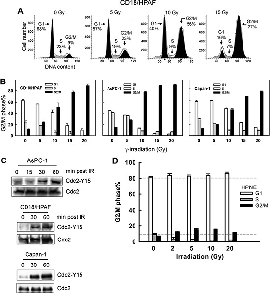
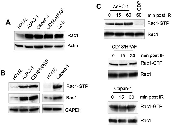
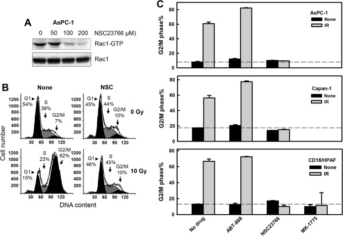
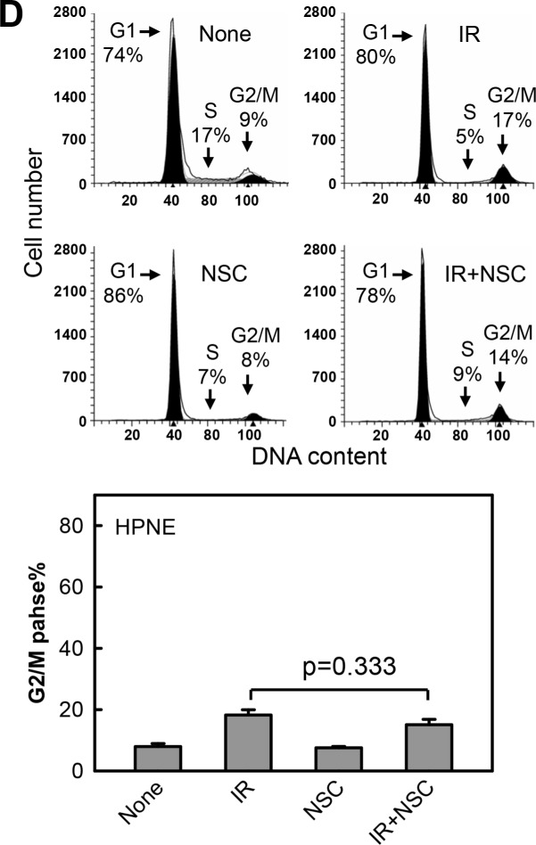
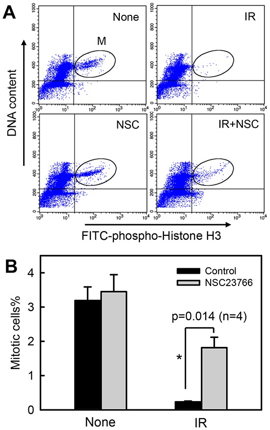
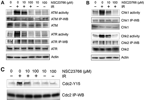
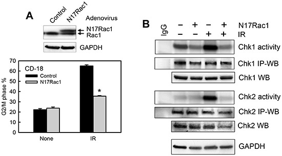
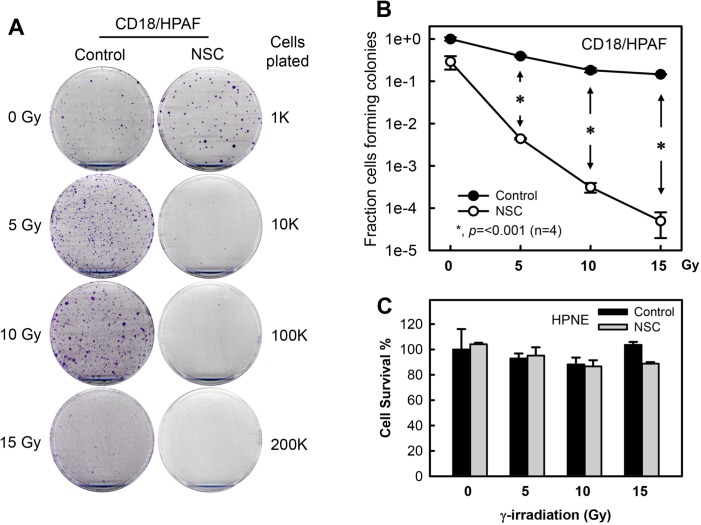
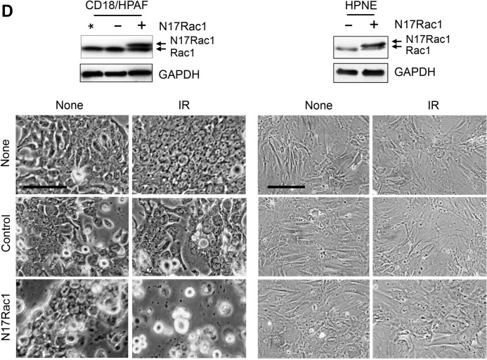
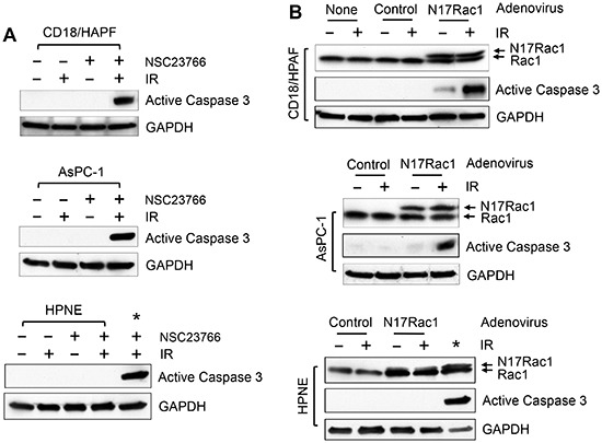
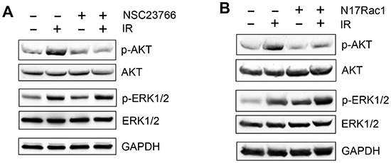
Radiation therapy is a staple treatment for pancreatic cancer. However, owing to the intrinsic radioresistance of pancreatic cancer cells, radiation therapy often fails to increase survival of pancreatic cancer patients. Radiation impedes cancer cells by inducing DNA damage, which can activate cell cycle checkpoints. Normal cells possess both a G1 and G2 checkpoint. However, cancer cells are often defective in G1 checkpoint due to mutations/alterations in key regulators of this checkpoint. Accordingly, our results show that normal pancreatic ductal cells respond to ionizing radiation (IR) with activation of both checkpoints whereas pancreatic cancer cells respond to IR with G2/M arrest only. Overexpression/hyperactivation of Rac1 GTPase is detected in the majority of pancreatic cancers. Rac1 plays important roles in survival and Ras-mediated transformation. Here, we show that Rac1 also plays a critical role in the response of pancreatic cancer cells to IR. Inhibition of Rac1 using specific inhibitor and dominant negative Rac1 mutant not only abrogates IR-induced G2 checkpoint activation, but also increases radiosensitivity of pancreatic cancer cells through induction of apoptosis. These results implicate Rac1 signaling in the survival of pancreatic cancer cells following IR, raising the possibility that this pathway contributes to the intrinsic radioresistance of pancreatic cancer.


Figures

Figure 1
Figure 1. IR induces G2/M cell cycle arrest and Cdc2-Y15 phosphorylation in pancreatic cancer cells
(A) Log-phase growing CD18/HPAF cells were exposed to increasing doses of IR, incubated for 24 h and analyzed for DNA content by FACS. Number of cells in G1, S and G2/M phases of the cell cycle are indicated. (B) Indicated pancreatic cancer cells were exposed to IR at the dose indicated, incubated for 24 h and analyzed for DNA content. Results depict the percentage of cells in G1 (white bars), S (gray bars) and G2/M (black bars) phases of the cell cycle and represent the mean±S.D. of two sets of experiments done in duplicates. (C) AsPC-1, CD18/HPAF and Capan-1 cells were exposed to 10 Gy IR, incubated for the indicated times and analyzed for Cdc2-Y15 phosphorylation as described in MATERIALS AND METHODS. As a control, levels of Cdc2 protein in cell lysates were assessed. (D) Normal human pancreatic ductal cells (HPNE) were exposed to IR at the doses indicated, incubated for 24 h and analyzed for DNA content by flow cytometry. The result depicts the percent cells in G1, S and G2/M phases of the cell cycle and is shown as the mean±S.D. of duplicate cell samples from two separate experiments.
Figure 2
Figure 2. Rac1 is overexpressed in pancreatic cancer cells
(A) Normal pancreatic ductal cells (HPNE) and pancreatic cancer cells (AsPC-1, Capan-1, CD18/HPAF and L3.6pl) were assessed for Rac1 protein expression by immunoblotting. (B) Indicated cells were analyzed for Rac1 activity (Rac1-GTP) as described in MATERIALS AND METHODS. As controls, protein levels of Rac1 (Rac1) and GAPDH (GAPDH) in cell lysates were measured. (C) AsPC-1, CD18/HPAF and Capan-1 cells were treated with 10 Gy IR and incubated for the indicated times and analyzed for the activity and level of Rac1.
Figure 3
Figure 3. Rac1 inhibition abrogates IR-induced G2/M cell cycle arrest in pancreatic cancer cells
(A) AsPC-1 cells were incubated for 1 h in the presence of NSC23766 at the indicated doses and analyzed for activity and level of Rac1. (B) AsPC-1 cells were incubated with 100 μM NSC23766 for 1 h, exposed to 10 Gy IR and incubated for 3 h post IR. The cells were washed, incubated in the absence of NSC23766 for 24 h and analyzed for DNA content by FACS. Number of cells in G1, S and G2/M phase of the cell cycle are indicated. (C) Indicated pancreatic cells were incubated for 1 h with ABT-888 (10 μM), NSC23766 (100 μM) and MK-1775 (3 μM), and exposed to 10 Gy IR. The cells were incubated for 3 h following IR, washed, incubated in the absence of drug for 24 h and analyzed for DNA content by FACS. Results depict the percentage of cells with 4N-DNA content (G2/M phase) and represent the mean±S.D. of two sets of experiments done in duplicates. (D) Upper panel: HPNE cells were incubated in the presence or absence of 100 μM NSC23766 for 1 h, exposed to 10 Gy IR, incubated for 24 h and analyzed for DNA content. Number of cells in G1, S and G2/M phase of the cell cycle are indicated. Lower panel: The result depicts the percentage of cells with 4N-DNA content (G2/M phase) and is shown as mean±S.D. of duplicate samples from two separate experiments.
Figure 4
Figure 4. Rac1 inhibition abrogates IR-induced G2/M checkpoint activation
CD18/HPAF cells were incubated for 1 h in the presence or absence of 100 μM NSC23766, treated with/without 10 Gy IR. After 2 h incubation following IR, the cells were analyzed by FACS for mitotic cells, which contain both 4N-DNA content and Histone H3-Ser10 phosphorylation [37]. (A) The histograms shown are representative FACS analyses for mitotic cells in samples treated with/without IR in the presence or absence of NSC23766. The location of mitotic cells in each sample is indicated (M). (B) The bar graph depicts the percentage of mitotic cells and is shown as mean±S.D. of duplicate samples from two set of experiments. *, significant difference from cells exposed to IR in the absence of NSC23766.
Figure 5
Figure 5. Rac1 inhibition abolishes IR-induced activation of both ATM and ATR signaling pathways
CD18/HPAF cells were treated with/without 10 Gy IR in the presence of NSC23766 at the indicated doses and incubated for 1 h at 37oC. (A) To assess ATR and ATM kinase activities, ATR and ATM were immunoprecipitated from the cell lysates using anti-ATR (N-19) and anti-ATM (2C1) antibodies respectively and assayed for relative kinase activity using recombinant p53 protein as substrate. (B) To measure Chk1 and Chk2 activity, Chk1 and Chk2 were immunoprecipitated from the cell lysates using anti-Chk1 (G-4) and anti-Chk2 (B-4) antibodies respectively and assayed for relative kinase activity using recombinant Cdc25C protein as substrate. As controls, protein levels of ATR, ATM, Chk1 and Chk2 in the immunoprecipitates (IP-WB) as well as in the cell lysates (WB) were assessed by immunoblotting. (C) Cdc2 was immunoprecipiated from the cell lysates using anti-Cdc2 (17) antibody and analyzed for Cdc2-Y15 phosphorylation and Cdc2 protein by immunoblotting.
Figure 6
Figure 6. Ectopic expression of N17Rac1 dominant mutant diminishes IR-induced G2/M checkpoint activation
(A) Upper panel: CD18/HPAF cells were transduced with adenoviral vector expressing N17Rac1 or control vector for 24 h and analyzed for Rac1 and GAPDH by immunoblotting. Lower panel: The transduced cells were treated with or without IR, incubated for 24 h and analyzed for DNA content by FACS. The result depicts the percentage of cells with 4N-DNA content and is shown as mean±S.D. of duplicate samples from two separate experiments. *, p< 0.001 (n=4), significant difference from the control vector transduced cells exposed to IR. (B) The transduced cells were treated with or without IR, incubated for 1 h and analyzed for the activities of Chk1 and Chk2 by kinase assay. As controls, protein levels of Chk1 and Chk2 in the immunoprecipitates (IP-WB) as well as in the cell lysates (WB) were assessed by immunoblotting. GAPDH protein in the cell lysates was assessed by Western blotting as a protein loading control.
Figure 7
Figure 7. Inhibition of Rac1 abrogates clonogenic survival of irradiated pancreatic cancer cells
(A) CD18/HPAF cells were exposed to increasing doses of IR in the presence or absence of 100 μM NSC23766 and incubated for 3 h. The cells were washed, incubated in regular medium for 14 days and assessed for numbers of colonies [63]. Representative sample dishes from the clonogenic assay are shown. (B) Number of colonies in the resulting samples (CD18/HPAF) was quantified using the ImageJ analytical program and the results are shown as mean±S.D. of two set of experiments done in duplicates. *, p=<0.001 (n=4), significant difference between the cells exposed to IR in the absence of NSC23766 and the cells exposed to IR in the presence of NSC23766. (C) HPNE cells were treated as described in (A). Cell survival in the resulting cell samples was quantified using the ImageJ analytical program and the results are shown as mean±S.D. of two set of experiments done in duplicates. (D) CD18/HPAF and HPNE cells were transduced with Ad.N17Rac1 (+) or Ad.Control (−) for 24 h. Upper panels: Western blot analysis of the indicated samples for Rac1 and GAPDH. *, un-transduced CD18/HPAF control cells. Lower panels: cells were treated with or without 10 Gy IR and incubated for additional 48 h. Cells were photographed using phase-contrast optics. Scale bars represent 100 μm.
Figure 8
Figure 8. Inhibition of Rac1 induces Caspase 3 activation in pancreatic cancer cells following IR
(A) The indicated cells were treated with/without 10 Gy IR in the presence or absence of 100 μM NSC23766 and incubated for 2 days. The cells were analyzed by immunoblotting for levels of activated Caspase 3 (p20) and GAPDH. *, positive control for caspase 3 activation: AsPC-1 cells treated with NSC23766 and IR. (B) The indicated cells were infected with Ad.N17Rac1 or Ad.Control for 24 h and exposed to 10 Gy IR or left non-irradiated. Following 24 h incubation, cells were examined by immunoblotting for levels of Rac1, activated Caspase 3 (p20) and GAPDH. *, positive control for caspase 3 activation: CD18/HPAF cells were transduced with Ad.N17Rac1 for 24 h, exposed to 10 Gy and incubated for 24 h.
Figure 9
Figure 9. Effect of Rac1 inhibition on IR-induced AKT and ERK1/2 phosphorylation
(A) In the presence or absence of 100 μM NSC23766, CD18/HPAF cells were treated with/without IR and analyzed for phosphorylation and level of AKT and ERK1/2 by immunoblotting. GAPDH was assessed as a protein loading control. (B) CD18/HPAF cell were infected with Ad.N17Rac1 or Ad.Control for 24 h and exposed to 10 Gy IR or un-irradiated. Following 1 h incubation post IR, the cells were examined for phosphorylation and level of AKT and ERK1/2. GAPDH was assessed as a protein loading control.

Similar articles

Cited by

References

    1. Callery MP, Chang KJ, Fishman EK, Talamonti MS, William Traverso L, Linehan DC. Pretreatment assessment of resectable and borderline resectable pancreatic cancer: expert consensus statement. Ann Surg Oncol. 2009;16:1727–1733. - PubMed
    1. Hazard L. The role of radiation therapy in pancreas cancer. Gastrointest Cancer Res. 2009;3:20–28. - PMC - PubMed
    1. Aref A, Berri R. Role of Radiation Therapy in the Management of Locally Advanced Pancreatic Cancer. Journal of Clinical Oncology. 2012;30:1564–1565. - PubMed
    1. Blackstock AW, Bernard SA, Richards F, Eagle KS, Case LD, Poole ME, Savage PD, Tepper JE. Phase I trial of twice-weekly gemcitabine and concurrent radiation in patients with advanced pancreatic cancer. J Clin Oncol. 1999;17:2208–2212. - PubMed
    1. Brade A, Brierley J, Oza A, Gallinger S, Cummings B, Maclean M, Pond GR, Hedley D, Wong S, Townsley C, Brezden-Masley C, Moore M. Concurrent gemcitabine and radiotherapy with and without neoadjuvant gemcitabine for locally advanced unresectable or resected pancreatic cancer: a phase I-II study. Int J Radiat Oncol Biol Phys. 2007;67:1027–1036. - PubMed

Publication types

MeSH terms