. 2014 Dec 15;5(23):11998-2015.
doi: 10.18632/oncotarget.2426.

The orthologous Tbx transcription factors Omb and TBX2 induce epithelial cell migration and extrusion in vivo without involvement of matrix metalloproteinases

Affiliations

The orthologous Tbx transcription factors Omb and TBX2 induce epithelial cell migration and extrusion in vivo without involvement of matrix metalloproteinases

Jie Shen et al. Oncotarget. .

Abstract

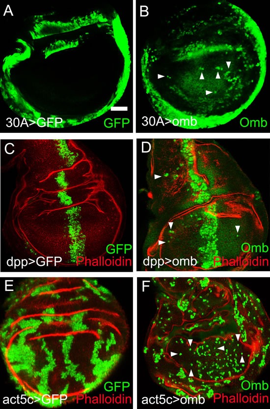
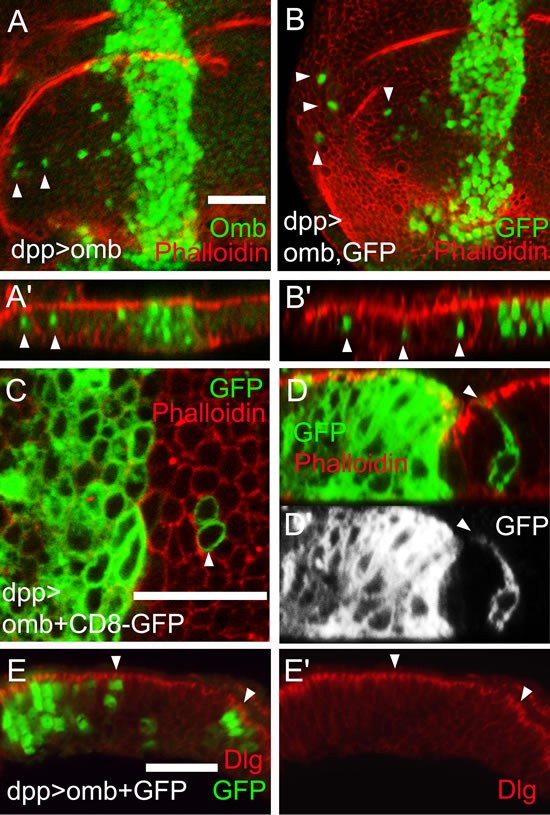
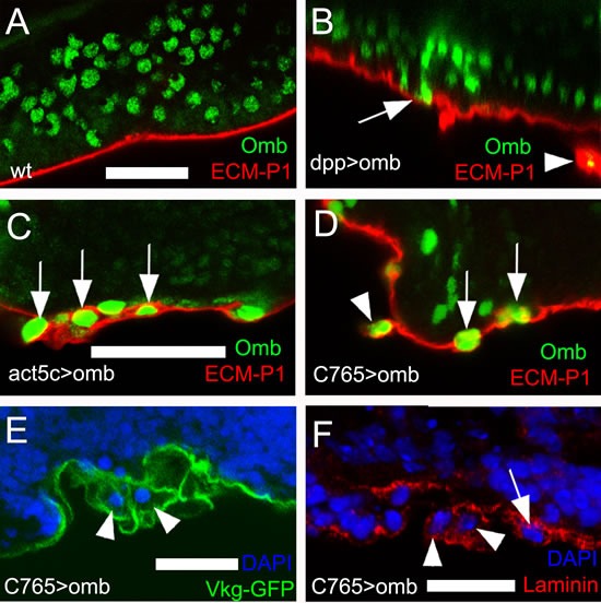
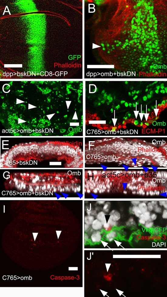
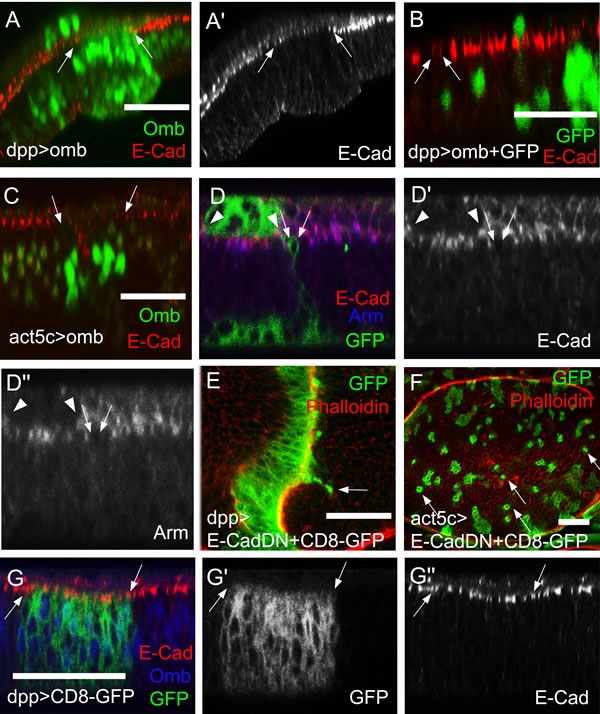
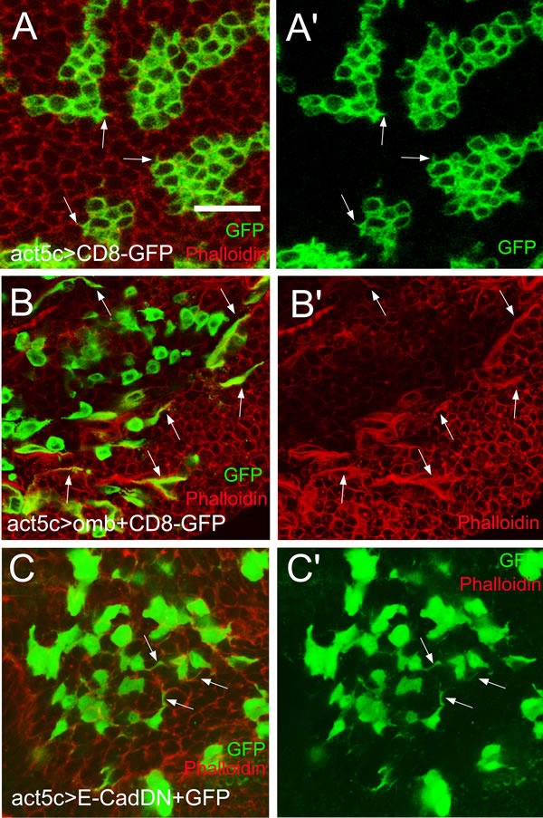
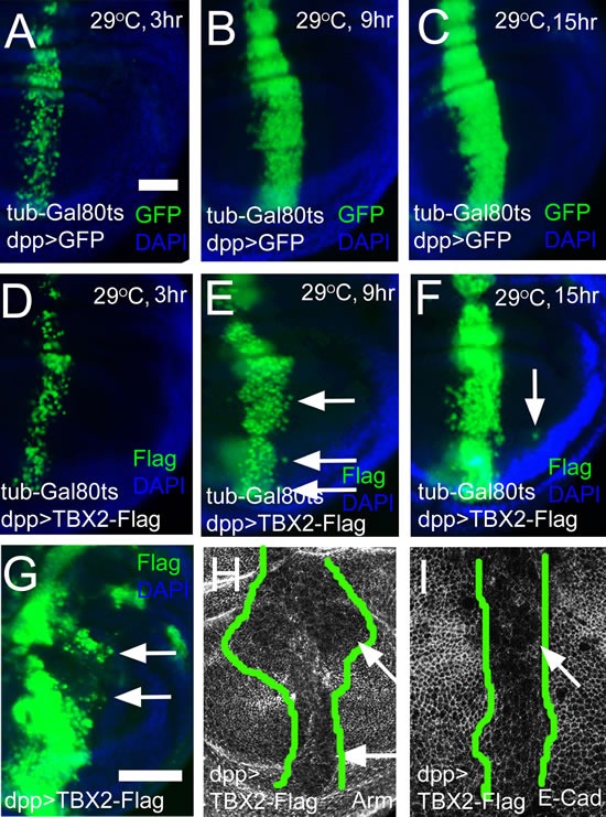
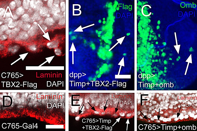
The transcription factors TBX2 and TBX3 are overexpressed in various human cancers. Here, we investigated the effect of overexpressing the orthologous Tbx genes Drosophila optomotor-blind (omb) and human TBX2 in the epithelium of the Drosophila wing imaginal disc and observed two types of cell motility. Omb/TBX2 overexpressing cells could move within the plane of the epithelium. Invasive cells migrated long-distance as single cells retaining or regaining normal cell shape and apico-basal polarity in spite of attenuated apical DE-cadherin concentration. Inappropriate levels of DE-cadherin were sufficient to drive cell migration in the wing disc epithelium. Omb/TBX2 overexpression and reduced DE-cadherin-dependent adhesion caused the formation of actin-rich lateral cell protrusions. Omb/TBX2 overexpressing cells could also delaminate basally, penetrating the basal lamina, however, without degradation of extracellular matrix. Expression of Timp, an inhibitor of matrix metalloproteases, blocked neither intraepithelial motility nor basal extrusion. Our results reveal an MMP-independent mechanism of cell invasion and suggest a conserved role of Tbx2-related proteins in cell invasion and metastasis-related processes.


Conflict of interest statement

Conflict of interest

The authors declare no conflict of interest.

Figures

Figure 1
Figure 1. Long distance migration of omb overexpressing cells in the Drosophila wing imaginal disc
In this and subsequent figures, wing imaginal discs were oriented anterior left and dorsal up. The developmental stages of the wing imaginal discs were middle to late third instar, unless indicated otherwise. x-y images were scanned at middle sections and were focussed on the wing pouch region, unless indicated otherwise. Scale bars are 50 μm. (A) Cells expressing GFP (green) in the 30A-Gal4 domain did not migrate out of their expression domain. In this and all subsequent panels, the symbol “>” denotes the connection between Gal4 driver and UAS regulated genes. Hence, 30A>GFP is short for 30A-Gal4, UAS-GFP. (B) Cells expressing Omb (green) in the 30A-Gal4 domain migrated into the central wing pouch. Cells derived from the 30-Gal4 domain were defined by high level Omb expression (intense green, arrowheads). The comparatively uniform and lower fluorescence intensity in the centre of the wing disc visualized the endogenous Omb expression. (C) Dpp>GFP cells (green) were confined to the dpp expression domain. (D) Cells overexpressing Omb (intense green) in the dpp-Gal4 domain migrated long distance away from the central expression stripe both into the anterior and posterior compartment (arrowheads). (E) Clonal cells expressing GFP (green) were clustered in groups. (F) Clones of cells overexpressing Omb (intense green) tended to disperse to the single cell level. In (C - F) wing discs were counterstained with Rhodamin phalloidin (red) which highlights folds in the epithelium.
Figure 2
Figure 2. Apparently normal cell shape of migrated cells
Cells overexpressing Omb (green) (A) or co-overexpressing omb with GFP (green) (B) in the dpp-Gal4 domain could migrate long distance to the periphery of the disc. In all x-z scans apical is up and anterior left. (A′ and B′) x-z scans of A and B. Nuclei of migrated cells had a wild-typic apico-basal position (arrowheads). (C and D) Cells co-overexpressing omb along with CD8-GFP (green) after crossing the A/P compartment boundary had wild-typic cell diameter (C) and apico-basal extension (D and D′). In A-D wing discs were counterstained with rhodamine phalloidin (red) highlighting cortical filamentous actin. (E) Cells that overexpressed omb (green) did not affect the subcellular distribution of Dlg (red), neither in the dpp expression domain (at the left end of panel) nor in migrated cells (arrowheads).
Figure 3
Figure 3. Omb overexpressing cells penetrate the ECM
ECM was stained by the fluorescent core-shell macromolecule ECM-P1 (red), Vkg-GFP (green), and anti-Laminin (red), respectively. All panels are x-z scans. (A) and (B), and (C) and (D) are at the same magnification, respectively. (A) ECM-P1 dye specifically stained the basement membrane of wild type wing disc. (B) Individual dpp>omb cells (green) apparently could penetrate the ECM (arrow). In such preparations, ECM-clad cells outside the basement membrane were observed (arrowhead). (C) Cells overexpressing Omb (green) derived from Act5C>omb clones could penetrate the ECM (arrows). (D-F) omb was overexpressed by the imaginal disc-specific driver C765-Gal4. Arrowheads and arrows indicate omb overexpressing cells penetrating the ECM (arrowheads) or located peripheral to the basement membrane (arrows).
Figure 4
Figure 4. JNK signalling and apoptosis are not required for omb-induced cell motility
(A) Expression of bsk[DN] did not elicit cell motility. Bsk[DN]-expressing cells are marked by co-expression of GFP (green). (B) Cells co-overexpressing omb and bsk[DN] in the dpp-Gal4 domain were still motile (arrowhead). (C) Clone cells co-overexpressing omb and bsk[DN] were dispersed to single cells (arrowheads). (D) - (H) and (J, J′) are x-z scans. (D) Cells co-overexpressing omb and bsk[DN] still invaded the basal ECM (arrows). (E) Ubiquitous expression of bsk[DN] did not cause basal delamination. (F) Disc-wide overexpression of omb induced extrusion of cells (arrowheads) which was not prevented by co-expression of bsk[DN] (G) or p35 (H). (I) Ubiquitous omb overexpression caused little caspase-3 activation (arrowheads). (J) Ubiquitous omb overexpression (C765>omb) induced basal invasion (arrows) not associated with anti-Caspase staining. A rare P-cas-3 positive cell is also contained in the picture (arrowhead). In (A), (B), and (E) - (H) discs were counterstained with rhodamine phalloidin (red).
Figure 5
Figure 5. DE-Cad and Arm levels are reduced in omb overexpressing cells
(A-D′) and (G-G″) are x-z scans. (A) When Omb (green) was overexpressed in the dpp-Gal4 domain, the DE-cad level (red) was attenuated (between arrows). (B) Migrated omb overexpressing cells (green) exhibited a reduced apical DE-cad level (arrows). The single green nucleus lying apical to the band of E-cad staining is in the peripodial membrane where dpp-Gal4 is also active. (C) When omb was expressed in clones, the DE-cadherin level was attenuated in the range of the clone. (D-D″) omb was co-expressed with CD8-GFP in clones (green). Single omb overexpressing and non-extruded cell had reduced apical DE-cad (red) and Arm (blue) levels. This also held for the clone in the periopodial epithelium outlined by arrowheads. (E) Cells expressing dominant negative DE-cad in the dpp-Gal4 domain could migrate across the A/P compartment boundary (arrow). (F) Clones of cells expressing dominant negative DE-cad (green) tended to disperse to the single cell level (arrows). Discs in (E) and (F) were counterstained with rhodamine phalloidin (red). (G) In a control wing imaginal disc expressing only the plasma membrane marker (CD8-GFP, green), E-cad expression (red) was uniform in- and outside the dpp-Gal4 expression domain, marked by arrows. The disc was counterstained against Omb (blue).
Figure 6
Figure 6. Actin-rich cellular protrusions in migratory omb overexpressing cells
(A and A′) Control CD8-GFP clones with short lateral protrusions (arrows). (B and B′) Omb-overexpressing cells, visualized by co-expression with the membrane marker CD8-GFP, were dispersed and frequently exhibited long lateral protrusions enriched in F-actin (arrows). (C and C′) Expressing a dominant negative form of DE-Cad induced long lateral protrusions (arrows). Filamentous actin was visualized by counterstaining with rhodamine phalloidin (red).
Figure 7
Figure 7. Expression of human TBX2 induces long distance migration and down-regulates adhesion junction molecules
(A-C) Time course of GFP expression (green) in the dpp-Gal4 domain. The hours after inactivation of Gal80ts by temperature upshift are indicated. (D-F) Time course of human TBX2-Flag (green) expression in the dpp-Gal4 domain. Nine hours after induction, TBX2 expressing cells had started to migrate across the A/P boundary (E, arrows). After 15 hours of induction, TBX2 cells were found at a larger distance from the dpp-Gal4 domain (F, arrow). (G) Constitutive expression of TBX2 in the dpp-Gal4 domain induced cells migration (arrows). In (A) - (G) discs were counterstained with DAPI (blue). (H and I) Apical sections showed the reduction of Arm (H) and E-Cad (I) in TBX2 expressing cells. The range of TBX2 expression is outlined by green lines based on the anti-Flag staining.
Figure 8
Figure 8. MMP activity is not required for intraepithelial motility and basal delamination induced by overexpression of TBX2 and Omb
In (A) and (D) - (F) nuclei are counterstained with DAPI (white); In (B) - (C) DAPI is blue. (A) Expressing TBX2 induced invasion and penetration of the basement membrane (arrows) leading to externalized cells coated with laminin (red). (B, C) Co-expressing Timp with TBX2 (B) or Omb (C) did not prevent long distance migration (arrows). (D) Wild type control in x-z scan. Wild-typic cell nuclei were not observed within the basement membrane of the epithelium. (E, F) Co-expressing Timp with TBX2 (E) or Omb (F) did not prevent basal delamination.

Similar articles

Cited by

References

    1. Hoek K, Rimm DL, Williams KR, Zhao H, Ariyan S, Lin A, Kluger HM, Berger AJ, Cheng E, Trombetta ES, Wu T, Niinobe M, Yoshikawa K, Hannigan GE, Halaban R. Expression profiling reveals novel pathways in the transformation of melanocytes to melanomas. Cancer Res. 2004;64(15):5270–5282. - PubMed
    1. Vance KW, Carreira S, Brosch G, Goding CR. Tbx2 is overexpressed and plays an important role in maintaining proliferation and suppression of senescence in melanomas. Cancer Res. 2005;65(6):2260–2268. - PubMed
    1. Boyd SC, Mijatov B, Pupo GM, Tran SL, Gowrishankar K, Shaw HM, Goding CR, Scolyer RA, Mann GJ, Kefford RF, Rizos H, Becker TM. Oncogenic B-RAF(V600E) Signaling Induces the T-Box3 Transcriptional Repressor to Repress E-Cadherin and Enhance Melanoma Cell Invasion. J Invest Dermatol. 2013;133(5):1269–1277. - PMC - PubMed
    1. Abrahams A, Parker MI, Prince S. The T-box transcription factor Tbx2: its role in development and possible implication in cancer. IUBMB Life. 2010;62(2):92–102. - PubMed
    1. Rowley M, Grothey E, Couch FJ. The role of Tbx2 and Tbx3 in mammary development and tumorigenesis. J Mammary Gland Biol Neoplasia. 2004;9(2):109–118. - PubMed

Publication types

MeSH terms

LinkOut - more resources