

TM4SF4 overexpression in radiation-resistant lung carcinoma cells activates IGF1R via elevation of IGF1
- PMID: 25344917
- PMCID: PMC4259440
- DOI: 10.18632/oncotarget.2450
TM4SF4 overexpression in radiation-resistant lung carcinoma cells activates IGF1R via elevation of IGF1
Abstract
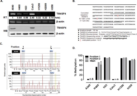
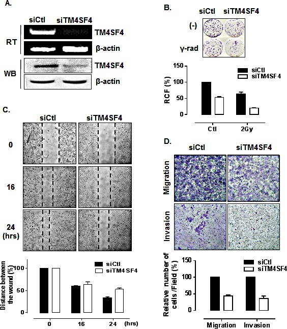
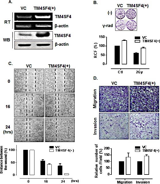
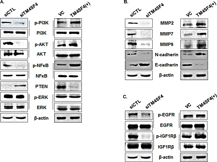
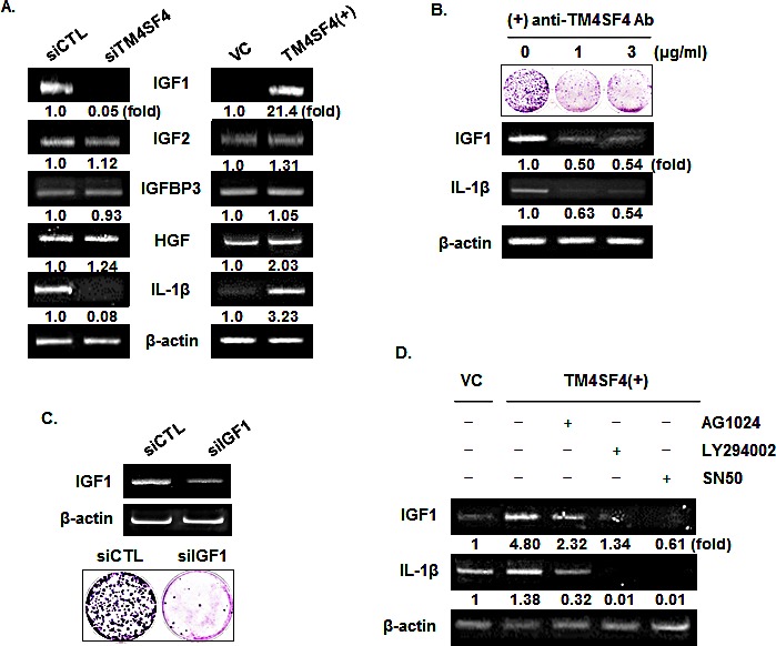
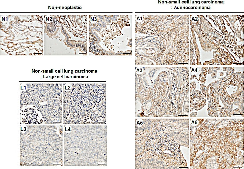
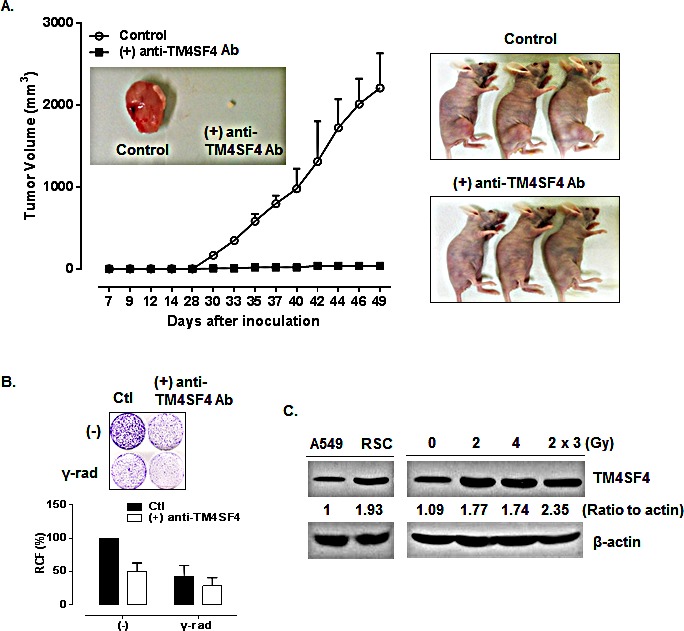
Transmembrane 4 L six family member 4 (TM4SF4) is a member of the tetraspanin L6 domain family. Other members of this family, TM4SF1 (also known as L6-Ag) and TM4SF5, have been shown to be upregulated in multiple tumors and involved in epithelial-to-mesenchymal transition and cell migration. However, unlike its homologs, little is known about TM4SF4. Here, we show that TM4SF4 was highly expressed in radiation-resistant lung adenocarcinoma cells, such as A549 and Calu-3 cells, and its expression activated cell growth, migration, and invasion. Overexpression of TM4SF4 in A549 cells increased the activation of PI3K, AKT, and NF-kappaB and the expression of PTEN. IGF1R was clearly activated by overexpression of TM4SF4, although EGFR was also slightly activated. TM4SF4 expression was correlated with the increased expression of IGF1, consequently resulting in IGF1R activation. Tumorigenic activity of TM4SF4 in lung adenocarcinoma cells was also demonstrated by xenograft assay; however, this activity was almost completely suppressed by treatment with anti-TM4SF4 antibody. Our results suggest that TM4SF4 overexpression in lung carcinoma cells results in resistance to radiotherapy via IGF1-induced IGF1R activation and blocking the activity of TM4SF4 using specific antibody can be a promising therapeutics against TM4SF4-overexpressing lung adenocarcinoma.
Figures
Similar articles
-
Three Members of Transmembrane-4-Superfamily, TM4SF1, TM4SF4, and TM4SF5, as Emerging Anticancer Molecular Targets against Cancer Phenotypes and Chemoresistance.Pharmaceuticals (Basel). 2023 Jan 11;16(1):110. doi: 10.3390/ph16010110. Pharmaceuticals (Basel). 2023. PMID: 36678607 Free PMC article. Review.
-
Bidirectional signaling between TM4SF5 and IGF1R promotes resistance to EGFR kinase inhibitors.Lung Cancer. 2015 Oct;90(1):22-31. doi: 10.1016/j.lungcan.2015.06.023. Epub 2015 Jul 2. Lung Cancer. 2015. PMID: 26190015
-
IGF1/IGF1R/STAT3 signaling-inducible IFITM2 promotes gastric cancer growth and metastasis.Cancer Lett. 2017 May 1;393:76-85. doi: 10.1016/j.canlet.2017.02.014. Epub 2017 Feb 20. Cancer Lett. 2017. PMID: 28223169
-
Fibulin-3-mediated inhibition of epithelial-to-mesenchymal transition and self-renewal of ALDH+ lung cancer stem cells through IGF1R signaling.Oncogene. 2014 Jul 24;33(30):3908-17. doi: 10.1038/onc.2013.373. Epub 2013 Sep 9. Oncogene. 2014. PMID: 24013232
-
Baicalein increases cisplatin sensitivity of A549 lung adenocarcinoma cells via PI3K/Akt/NF-κB pathway.Biomed Pharmacother. 2017 Jun;90:677-685. doi: 10.1016/j.biopha.2017.04.001. Epub 2017 Apr 14. Biomed Pharmacother. 2017. PMID: 28415048 Review.
Cited by
-
mRNA and methylation profiling of radioresistant esophageal cancer cells: the involvement of Sall2 in acquired aggressive phenotypes.J Cancer. 2017 Feb 25;8(4):646-656. doi: 10.7150/jca.15652. eCollection 2017. J Cancer. 2017. PMID: 28367244 Free PMC article.
-
Three Members of Transmembrane-4-Superfamily, TM4SF1, TM4SF4, and TM4SF5, as Emerging Anticancer Molecular Targets against Cancer Phenotypes and Chemoresistance.Pharmaceuticals (Basel). 2023 Jan 11;16(1):110. doi: 10.3390/ph16010110. Pharmaceuticals (Basel). 2023. PMID: 36678607 Free PMC article. Review.
-
Molecular Signatures of the Insulin-like Growth Factor 1-mediated Epithelial-Mesenchymal Transition in Breast, Lung and Gastric Cancers.Int J Mol Sci. 2018 Aug 15;19(8):2411. doi: 10.3390/ijms19082411. Int J Mol Sci. 2018. PMID: 30111747 Free PMC article. Review.
-
miR-30 Family Reduction Maintains Self-Renewal and Promotes Tumorigenesis in NSCLC-Initiating Cells by Targeting Oncogene TM4SF1.Mol Ther. 2018 Dec 5;26(12):2751-2765. doi: 10.1016/j.ymthe.2018.09.006. Epub 2018 Sep 13. Mol Ther. 2018. PMID: 30301667 Free PMC article.
-
Clinical significance of TM4SF1 as a tumor suppressor gene in gastric cancer.Cancer Med. 2018 Jun;7(6):2592-2600. doi: 10.1002/cam4.1494. Epub 2018 Apr 17. Cancer Med. 2018. PMID: 29665316 Free PMC article.
References
-
- Muller-Pillasch F, Wallrapp C, Lacher U, Friess H, Buchler M, Adler G, Gress TM. Identification of a new tumour-associated antigen TM4SF5 and its expression in human cancer. Gene. 1998;208(1):25–30. - PubMed
-
- Kaneko R, Tsuji N, Kamagata C, Endoh T, Nakamura M, Kobayashi D, Yagihashi A, Watanabe N. Amount of expression of the tumor-associated antigen L6 gene and transmembrane 4 superfamily member 5 gene in gastric cancers and gastric mucosa. The American journal of gastroenterology. 2001;96(12):3457–3458. - PubMed
Publication types
MeSH terms
Substances
LinkOut - more resources
Full Text Sources
Other Literature Sources
Medical
Molecular Biology Databases
Research Materials
Miscellaneous
