. 2015;61(1):13-9.
doi: 10.1262/jrd.2014-098. Epub 2014 Oct 25.

Naïve-like conversion enhances the difference in innate in vitro differentiation capacity between rabbit ES cells and iPS cells

Affiliations

Naïve-like conversion enhances the difference in innate in vitro differentiation capacity between rabbit ES cells and iPS cells

Kimiko Honsho et al. J Reprod Dev. 2015.

Abstract

Quality evaluation of pluripotent stem cells using appropriate animal models needs to be improved for human regenerative medicine. Previously, we demonstrated that although the in vitro neural differentiating capacity of rabbit induced pluripotent stem cells (iPSCs) can be mitigated by improving their baseline level of pluripotency, i.e., by converting them into the so-called "naïve-like" state, the effect after such conversion of rabbit embryonic stem cells (ESCs) remains to be elucidated. Here we found that naïve-like conversion enhanced the differences in innate in vitro differentiation capacity between ESCs and iPSCs. Naïve-like rabbit ESCs exhibited several features indicating pluripotency, including the capacity for teratoma formation. They differentiated into mature oligodendrocytes much more effectively (3.3-7.2 times) than naïve-like iPSCs. This suggests an inherent variation in differentiation potential in vitro among PSC lines. When naïve-like ESCs were injected into preimplantation rabbit embryos, although they contributed efficiently to forming the inner cell mass of blastocysts, no chimeric pups were obtained. Thus, in vitro neural differentiation following naïve-like conversion is a promising option for determining the quality of PSCs without the need to demonstrate chimeric contribution. These results provide an opportunity to evaluate which pluripotent stem cells or treatments are best suited for therapeutic use.


Figures
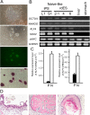
Fig. 1.
Fig. 1.
Characterization of naïve-like rabbit ESCs. (A) Colony morphology of primed-state rabbit ESCs (rdES2-1 cell line) (top panel), naïve-like converted ESCs (upper middle panel), expressing hOCT3/4-IRES-GFP (lower middle panel) and demonstrating alkaline phosphatase activity (bottom panel). Scale bar = 200 μm. (B) RT-PCR analysis of pluripotent marker genes in naïve-like converted liver-derived iPS cells (iPS-L1), stomach-derived iPS cells (iPS-S1), ES cell lines (2-1, 4 and 6) and somatic cells (liver, stomach). (C) Quantitative real-time RT-PCR analysis of KLF4 and KLF5 expression in primed (closed bar) and naïve-like (open bar) ESCs (rdES2-1 cells). (D) Naïve-like converted ESCs were able to form teratomas containing tissues originating from the three primary germ layers: epithelium with Goblet cells (left panel; endoderm), bone (middle panel; mesoderm) and epidermis (right panel; ectoderm).
Fig. 2.
Fig. 2.
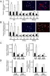
Evaluation of the capacity for differentiation into oligodendrocytes among primed and naïve-like ESCs/iPSCs. Immunohistochemical detection of oligodendrocytes was evaluated using anti-O1 antibody (red) (A) and anti-O1 and anti-CNPase merged (orange) (B) signals (arrowheads) before (P) and after (N) naïve-like conversion of PSC lines. Mature oligodendrocytes with dendrites (arrows) were derived from naïve-like rdES2-1 cells (right panels). Quantification of the neural differentiation index of primed (P; closed bar) and naïve-like (N; open bar) rabbit ESC lines (rdES2-1, rdES4 and rdES6) and rabbit iPSC lines (liver-derived iPS-L1, iPS-L2 and iPS-L3 cells and stomach-derived iPS-S1, iPS-S2 and iPS-S3 cells). These signals were normalized to DAPI DNA staining signals. Data are shown as the mean ± SE (n = 3–7). *P < 0.05. Scale bar =100 μm. (C) Relative neural differentiation efficiencies of ESC lines and iPSC lines before (P; closed bar) and after (N; open bar) naïve-like conversion. Immunostaining signals for O1 (left graph) and O1/CNPase merged (right graph) from ESCs (rdES2-1, rdES4 and rdES6), liver-derived iPSCs (iPS-L1, iPS-L2 and iPS-L3) and stomach-derived iPSCs (iPS-S1, iPS-S2 and iPS-S3) have been averaged. Data are shown as the mean ± SE (n = 9–21). *P < 0.05. (D) The relative differentiation efficiencies before (P; closed bar) and after (N; open bar) naïve-like conversion of ESCs and iPSCs are shown. Immunostaining signals for O1 (left graph) and O1/CNPase merged (right graph) from ES cells (rdES2-1, rdES4 and rdES6) were normalized against liver-derived iPSCs (iPS-L1, iPS-L2 and iPS-L3) or stomach-derived iPSCs (iPS-S1, iPS-S2 and iPS-S3).
Fig. 3.
Fig. 3.
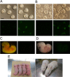
Chimeric contribution of naïve-like ESCs in reconstructed embryos and pups. (A) EF1α–OCT3/4–GFP expression in rabbit blastocysts 48 h after an injection into 8-cell rabbit embryos of primed-state rdES2-1 cells (left panels) and naïve-like rdES2-1 cells (right panels). When primed-state ESCs were injected, putatively differentiated ESCs with almost no hOCT3/4–GFP expression did not contribute to the ICM of blastocysts. Scale bar = 100 μm. (B) EF1α–OCT3/4–GFP expression in interspecific (rabbit and mouse) blastocysts 48 h after injection into 8-cell mouse embryos of primed-state rdES2-1 cells (left panels) and naïve-like rdES2-1 cells (right panels). (C, D) Detection of chimeric contribution by GFP signals in a reconstructed rabbit embryo (15.5 dpc) (C) and interspecific (rabbit and mouse) embryo (11.5 dpc) (D). No GFP signal was observed (right panels). (E) Detection of chimeric contribution by black coat color. No chimeric contribution was observed in the pups (skin; left panel) or does (hair; right panel).

Similar articles

Cited by

References

    1. Honda A, Hirose M, Inoue K, Ogonuki N, Miki H, Shimozawa N, Hatori M, Shimizu N, Murata T, Hirose M, Katayama K, Wakisaka N, Miyoshi H, Yokoyama KK, Sankai T, Ogura A. Stable embryonic stem cell lines in rabbits: potential small animal models for human research. Reprod Biomed Online 2008; 17: 706–715. - PubMed
    1. Honda A, Hirose M, Hatori M, Matoba S, Miyoshi H, Inoue K, Ogura A. Generation of induced pluripotent stem cells in rabbits: potential experimental models for human regenerative medicine. J Biol Chem 2010; 285: 31362–31369. - PMC - PubMed
    1. Graur D, Duret L, Gouy M. Phylogenetic position of the order Lagomorpha (rabbits, hares and allies). Nature 1996; 379: 333–335. - PubMed
    1. Shiomi M, Ito T, Yamada S, Kawashima S, Fan J. Correlation of vulnerable coronary plaques to sudden cardiac events. Lessons from a myocardial infarction-prone animal model (the WHHLMI rabbit). J Atheroscler Thromb 2004; 11: 184–189. - PubMed
    1. Weekers F, Van Herck E, Coopmans W, Michalaki M, Bowers CY, Veldhuis JD, Van den Berghe G. A novel in vivo rabbit model of hypercatabolic critical illness reveals a biphasic neuroendocrine stress response. Endocrinology 2002; 143: 764–774. - PubMed

Publication types

Substances