Review
. 2014 Nov;24(6):631-53.
doi: 10.1111/bpa.12196.

The 18 kDa translocator protein, microglia and neuroinflammation

Affiliations
Review

The 18 kDa translocator protein, microglia and neuroinflammation

Guo-Jun Liu et al. Brain Pathol. 2014 Nov.

Abstract

The 18 kDa translocator protein (TSPO), previously known as the peripheral benzodiazepine receptor, is expressed in the injured brain. It has become known as an imaging marker of "neuroinflammation" indicating active disease, and is best interpreted as a nondiagnostic biomarker and disease staging tool that refers to histopathology rather than disease etiology. The therapeutic potential of TSPO as a drug target is mostly based on the understanding that it is an outer mitochondrial membrane protein required for the translocation of cholesterol, which thus regulates the rate of steroid synthesis. This pivotal role together with the evolutionary conservation of TSPO has underpinned the belief that any loss or mutation of TSPO should be associated with significant physiological deficits or be outright incompatible with life. However, against prediction, full Tspo knockout mice are viable and across their lifespan do not show the phenotype expected if cholesterol transport and steroid synthesis were significantly impaired. Thus, the "translocation" function of TSPO remains to be better substantiated. Here, we discuss the literature before and after the introduction of the new nomenclature for TSPO and review some of the newer findings. In light of the controversy surrounding the function of TSPO, we emphasize the continued importance of identifying compounds with confirmed selectivity and suggest that TSPO expression is analyzed within specific disease contexts rather than merely equated with the reified concept of "neuroinflammation."

Keywords: PBR111; PET; PK11195; TSPO; microglia; neuroinflammation.


Figures

Figure 1
Figure 1
Figure 1 shows the increase in microglia literature. There is a steep rise from around 1995 in the overall publication activity under the heading of “microglia” that coincides with emergence of the themes “microglia and neuroinflammation” (7307 publications), “microglia and brain imaging” (3926 publications), “microglia and PET” (1094 publications) and “microglia and TSPO” (653 publications). The arrows in the upper, larger panel indicate the appearance of the first articles (1997–2000) retrieved under the specified search terms.
Figure 2
Figure 2
Adapted from 16. The transition of parenchymal microglia from their normal state to an activated state is accompanied by the de novo expression of binding sites for (R)‐PK11195, the prototypical ligand originally used to describe the presence and function of TSPO 100. Transformation of microglia into ameboid cells, such as seen after injuries involving neuronal cell death 20, does not appear to lead to any further increase in TSPO expression as measured by binding of (R)‐PK11195. This implies that the dynamic range of signals based on the expression of TSPO is best exploited at the point of transition from resting to activated, but not ameboid, microglia.
Figure 3
Figure 3
Adapted from 17. (A) The blood–brain barrier (BBB) separates the brain from the peripheral immune system. TSPO expression in the brain is low to absent. (B) Pathologies causing disruption of the BBB allow the influx of cells of the peripheral immune system. TSPO expression may be due to activated microglia or invading blood‐borne macrophages. (C) Noninflammatory neuronal injuries without obvious BBB damage, such as after peripheral nerve lesion, evoke microglial responses locally and in projection areas. TSPO expression occurs throughout a network of neural tracts. (D) The presence of activated microglia can confer regional “immune alertness” with the secondary site‐directed recruitment of, for example, circulating T‐lymphocytes. The neuronally triggered glial response may evolve toward a delayed inflammatory response.
Figure 4
Figure 4
From 37. The 3D [11C](R)‐PK11195 binding potential map projected onto the magnetic resonance image (MRI) of a patient with herpes encephalitis indicates how the inflammation in the temporal lobe and hippocampus (red) is associated with TSPO expression that tracks throughout the limbic system beyond the focal injury. This is likely to be due to Wallerian degeneration along the affected neural tract system (blue = anterior cingulate; green = thalamus and brainstem; purple = insular cortex; yellow = orbitofrontal gyri; the MRI subtraction image in the right panel shows the volume losses, including the enlargement of the ventricles (v) of the destructive tissue pathology).
Figure 5
Figure 5
Clinical trials listed under “neuroinflammation” on clinicaltrials.gov as of August 1, 2014.
Figure 6
Figure 6
Top 10 SCOPUS research areas by citation rates of TSPO‐related articles. Neuroscience is one of the leading areas for TSPO research.
Figure 7
Figure 7
(A) Publication of articles relating to TSPO per year demonstrates two waves of publication, roughly corresponding to protein/binding site characterization and later application of ligands for imaging and preclinical studies. (B) Citation of articles relating to TSPO have increased in the wake of potential applications of ligands.
Figure 8
Figure 8
The topology of TSPO in the membrane, with amino acids involved in the binding of Ro5‐4863, PK11195, Alpidem and the CRAC domain highlighted.
Figure 9
Figure 9
Structures of diazepam and Ro5‐4864.
Figure 10
Figure 10
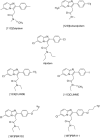
Ligands from the imidazopyridine class.
Figure 11
Figure 11
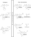
The major metabolites of the [123I]CLINDE, [18F]PBR102, [18F]PBR111 and [18F]DPA714.
Figure 12
Figure 12
Structure of PBR170.

References

    1. Adam D (2002) Citation analysis: the counting house. Nature 415:726–729. - PubMed
    1. Alenfall J, Kant R, Batra S (1998) Cytotoxic effects of 125I‐labeled PBZr ligand PK 11195 in prostatic tumor cells: therapeutic implications. Cancer Lett 134:187–192. - PubMed
    1. Anholt RR, De Souza EB, Kuhar MJ, Snyder SH (1985) Depletion of peripheral‐type benzodiazepine receptors after hypophysectomy in rat adrenal gland and testis. Eur J Pharmacol 110:41–46. - PubMed
    1. Anholt RR, De Souza EB, Oster‐Granite ML, Snyder SH (1985) Peripheral‐type benzodiazepine receptors: autoradiographic localization in whole‐body sections of neonatal rats. J Pharmacol Exp Ther 233:517–526. - PubMed
    1. Anholt RR, Pedersen PL, De Souza EB, Snyder SH (1986) The peripheral‐type benzodiazepine receptor. Localization to the mitochondrial outer membrane. J Biol Chem 261:576–583. - PubMed

LinkOut - more resources