

Genomewide analysis of rat periaqueductal gray-dorsal horn reveals time-, region- and frequency-specific mRNA expression changes in response to electroacupuncture stimulation
- PMID: 25346229
- PMCID: PMC4209446
- DOI: 10.1038/srep06713
Genomewide analysis of rat periaqueductal gray-dorsal horn reveals time-, region- and frequency-specific mRNA expression changes in response to electroacupuncture stimulation
Abstract
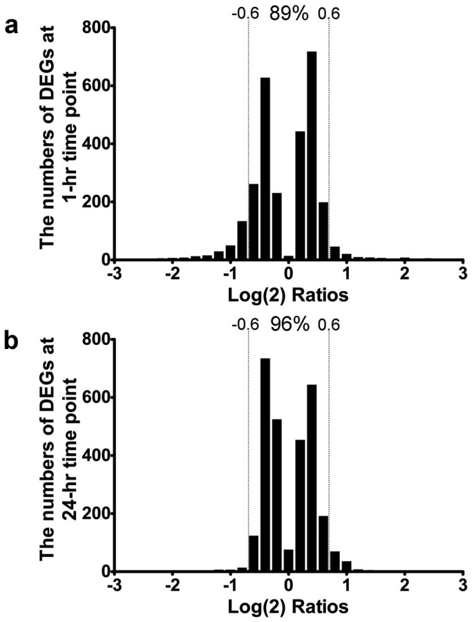
Electroacupuncture (EA) has been widely applied for illness prevention, treatment or rehabilitation in the clinic, especially for pain management. However, the molecular events that induce these changes remain largely uncharacterized. The periaqueductal gray (PAG) and the spinal dorsal horn (DH) have been verified as two critical regions in the response to EA stimulation in EA analgesia. In this study, a genetic screen was conducted to delineate the gene expression profile in the PAG-DH regions of rats to explore the molecular events of the analgesic effect induced by low-frequency (2-Hz) and high-frequency (100-Hz) EAs. Microarray analysis at two different time points after EA stimulation revealed time-, region- and frequency-specific gene expression changes. These expression differences suggested that modulation of neural-immune interaction in the central nervous system played an important role during EA analgesia. Furthermore, low-frequency EA could regulate gene expression to a greater degree than high-frequency EA. Altogether, the present study offers, for the first time, a characterized transcriptional response pattern in the PAG-DH regions followed by EA stimulation and, thus, provides a solid experimental framework for future in-depth analysis of the mechanisms underlying EA-induced effects.
Figures


References
-
- NIH Consensus Conference. Acupuncture. JAMA 280, 1518–1524 (1998). - PubMed
-
- Zhao Z. Q. Neural mechanism underlying acupuncture analgesia. Prog Neurobiol 85, 355–375 (2008). - PubMed
-
- Han J. S. Acupuncture: neuropeptide release produced by electrical stimulation of different frequencies. Trends Neurosci 26, 17–22 (2003). - PubMed
-
- Silva J. R., Silva M. L. & Prado W. A. Analgesia induced by 2- or 100-Hz electroacupuncture in the rat tail-flick test depends on the activation of different descending pain inhibitory mechanisms. J Pain 12, 51–60 (2011). - PubMed
-
- Hui K. K. et al. The integrated response of the human cerebro-cerebellar and limbic systems to acupuncture stimulation at ST 36 as evidenced by fMRI. Neuroimage 27, 479–496 (2005). - PubMed
Publication types
MeSH terms
LinkOut - more resources
Full Text Sources
Other Literature Sources
Molecular Biology Databases
