Review
. 2015 Mar-Apr;6(2):225-42.
doi: 10.1002/wrna.1269. Epub 2014 Oct 27.

An overview of pre-ribosomal RNA processing in eukaryotes

Affiliations
Review

An overview of pre-ribosomal RNA processing in eukaryotes

Anthony K Henras et al. Wiley Interdiscip Rev RNA. 2015 Mar-Apr.

Abstract

Ribosomal RNAs are the most abundant and universal noncoding RNAs in living organisms. In eukaryotes, three of the four ribosomal RNAs forming the 40S and 60S subunits are borne by a long polycistronic pre-ribosomal RNA. A complex sequence of processing steps is required to gradually release the mature RNAs from this precursor, concomitant with the assembly of the 79 ribosomal proteins. A large set of trans-acting factors chaperone this process, including small nucleolar ribonucleoparticles. While yeast has been the gold standard for studying the molecular basis of this process, recent technical advances have allowed to further define the mechanisms of ribosome biogenesis in animals and plants. This renewed interest for a long-lasting question has been fueled by the association of several genetic diseases with mutations in genes encoding both ribosomal proteins and ribosome biogenesis factors, and by the perspective of new anticancer treatments targeting the mechanisms of ribosome synthesis. A consensus scheme of pre-ribosomal RNA maturation is emerging from studies in various kinds of eukaryotic organisms. However, major differences between mammalian and yeast pre-ribosomal RNA processing have recently come to light.


Figures

Figure 1
Figure 1
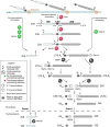
Pre-ribosomal RNA (rRNA) processing in yeast Saccharomyces cerevisiae. The majority of the nascent transcripts are cleaved co-transcriptionally at sites A0, A1, and A2, yielding the 20S and 27S-A2 pre-rRNAs (green). Alternatively, the full-length 35S pre-rRNA is processed post-transcriptionally (red). After elimination of the 5′-ETS and cleavage at site A2, maturation of the 18S rRNA 3′ end from the 20S pre-rRNA requires a single endonucleolytic cleavage step by Nob1p, which takes place in the cytoplasm after nuclear export of the pre-40S particle. Maturation of the large subunit follows two pathways, which yield two versions of the 5.8S rRNA 5′ end. The major pathway produces a short form by endonucleolytic cleavage of the 27S-A2 pre-rRNA at site A3 by RNase MRP and subsequent exonucleolytic processing of the 27S-A3 pre-rRNA by Rat1p and Rrp17p. Alternatively, the 27S-A2 pre-rRNA is cleaved at site B1L, yielding a long form of the 5.8S rRNA. Maturation of the 5.8S rRNA 3′ end is completed in the cytoplasm by exonuclease Ngl2p.
Figure 2
Figure 2
Pre-ribosomal RNA (rRNA) processing in mammalian cells. The pre-rRNA processing presented here combines data from studies in human and murine cells. The nomenclature refers to human cells,; the corresponding nomenclature in mouse is indicated in Figure 3. Alternative cleavage sequences are depicted in different colors. Short-lived precursors are represented with dotted lines. Cleavage of the 45S pre-rRNA can either start in the 5′-ETS (red) or in the ITS1 (green), which defines two pathways. If cleavage of the 5′-ETS occurs first (41S pre-rRNA), subsequent cleavage in the ITS1 takes place either at site 2 or at site E (purple). Initial cleavage at site 2 is the major pathway in HeLa cells, considering the abundance of the 30S pre-rRNA relative to the 41S. In mouse cell lines, the 36S pre-rRNA is readily detected. The endonuclease NOB1 is necessary for maturation of the 3′ end of the 18S-E pre-rRNA in the cytoplasm. Formation of the long and short 5′ ends of the 5.8S rRNA is not fully documented in mammalian cells. The 5.8S rRNA 3′-end maturation pathway primarily involves exonucleases,– but the 7S pre-rRNA was also proposed to result from endonucleolytic cleavage of the 12S pre-rRNA (site 4a in mouse, see Figure 3)., It has not been formally demonstrated that final maturation of the 6S pre-rRNA takes place in the cytoplasm in mammalian cells, but this was shown in Xenopus laevis and Saccharomyces cerevisiae.
Figure 3
Figure 3
The different pre-rRNAs detected in human and murine cells and their nomenclature.,,,,,,,, (a) Definition of the major pre-ribosomal RNAs (rRNAs). Arrowheads indicate the endonucleolytic cleavage sites. The yellow box corresponds to a highly conserved domain in ITS1 among mammals. Its boundaries define the 5′ end of the 36S-C and the 3′ end of the 21S-C, suggesting that exonucleolytic trimming of these extremities is stopped by a secondary structure and/or a protein complex anchored on this region. The 36S-C and the 30S+1/34.5S species are examples of precursors that are only detected upon perturbation of ribosome biogenesis. (b) Map of the processing sites in the human (orange) and murine (blue) pre-rRNAs. Arrowheads indicate the endonucleolytic cleavage sites. The nucleotides correspond to the residue located 5′ to the cleavage site. The extremities of the 18S, 5.8S, and 28S rRNAs are indicated in bold letters. The mapping of these sites is partly discussed in the text and was extensively reviewed by Mullineux and Lafontaine. Human site E was determined by primer extension and 3′-RACE. Localization of site 4b in mouse was mapped by primer extension. The human sites equivalent to sites 3 and 4a in mouse have not been formally designated. Details on site 4a are given in the text. The numbering of the nucleotides refers to GenBank sequences U13369.1 (human rDNA) and BK000964.3 (mouse rDNA).
Figure 4
Figure 4
Role of the ribosomal proteins in formation of the 40S subunit. (a) Secondary structure of the human 18S rRNA (http://apollo.chemistry.gatech.edu/RibosomeGallery/). Four topological domains can be distinguished, which form distinct features in the 3D structure: the body and the platform outlined in green, and the head outlined in purple. (b) Two functional groups in RPS proteins. A first class of RPSs in human cells is strictly required for initiating cleavage at sites A0, 1, and E. These ‘initiation RPSs’ or i-RPSs (blue) are associated with the 5′ part of the RNA (body, platform, and back of the head). The ‘progression RPSs’ or p-RPSs (orange) are subsequently required for efficient processing at sites 1, E, or 3; they assemble on or around the head, consistent with a delay in the formation of this structure with respect to the body and the platform. Consistent data were found in yeast., The figure shows the position of these proteins in the structure of the human 40S subunit solved by cryo-electron microscopy (PDB 3J3A). The 18S rRNA appears in beige.

References

    1. Klinge S, Voigts-Hoffmann F, Leibundgut M, Ban N. Atomic structures of the eukaryotic ribosome. Trends Biochem Sci. 2012;37:189–198. - PubMed
    1. Yusupova G, Yusupov M. High-resolution structure of the eukaryotic 80S ribosome. Annu Rev Biochem. 2014;83:467–486. - PubMed
    1. Warner JR. The economics of ribosome biosynthesis in yeast. Trends Biochem Sci. 1999;24:437–440. - PubMed
    1. Hernandez-Verdun D, Roussel P, Thiry M, Sirri V, Lafontaine DL. The nucleolus: structure/function relationship in RNA metabolism. WIREs RNA. 2010;1:415–431. - PubMed
    1. Babu KA, Verma RS. Structural and functional aspects of nucleolar organizer regions (NORs) of human chromosomes. Int Rev Cytol. 1985;94:151–176. - PubMed