Review
. 2014 Oct 9:8:293.
doi: 10.3389/fncel.2014.00293. eCollection 2014.

Motor neurons and the generation of spinal motor neuron diversity

Affiliations
Review

Motor neurons and the generation of spinal motor neuron diversity

Nicolas Stifani. Front Cell Neurosci. .

Abstract

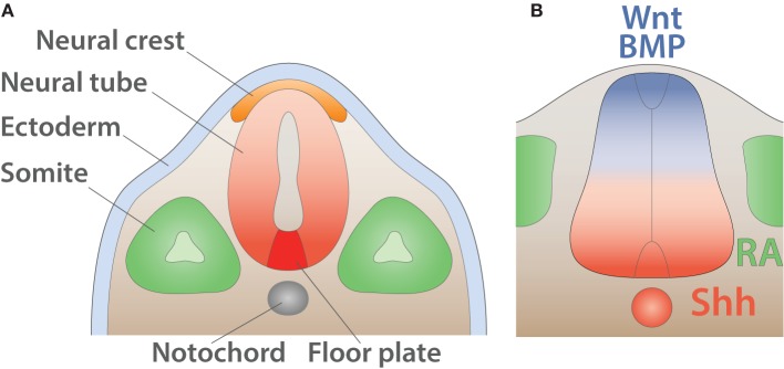
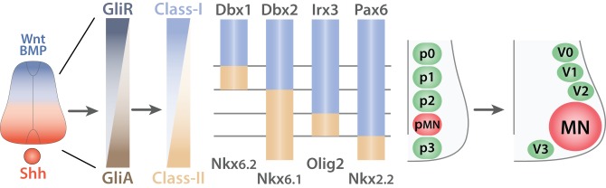
Motor neurons (MNs) are neuronal cells located in the central nervous system (CNS) controlling a variety of downstream targets. This function infers the existence of MN subtypes matching the identity of the targets they innervate. To illustrate the mechanism involved in the generation of cellular diversity and the acquisition of specific identity, this review will focus on spinal MNs (SpMNs) that have been the core of significant work and discoveries during the last decades. SpMNs are responsible for the contraction of effector muscles in the periphery. Humans possess more than 500 different skeletal muscles capable to work in a precise time and space coordination to generate complex movements such as walking or grasping. To ensure such refined coordination, SpMNs must retain the identity of the muscle they innervate. Within the last two decades, scientists around the world have produced considerable efforts to elucidate several critical steps of SpMNs differentiation. During development, SpMNs emerge from dividing progenitor cells located in the medial portion of the ventral neural tube. MN identities are established by patterning cues working in cooperation with intrinsic sets of transcription factors. As the embryo develop, MNs further differentiate in a stepwise manner to form compact anatomical groups termed pools connecting to a unique muscle target. MN pools are not homogeneous and comprise subtypes according to the muscle fibers they innervate. This article aims to provide a global view of MN classification as well as an up-to-date review of the molecular mechanisms involved in the generation of SpMN diversity. Remaining conundrums will be discussed since a complete understanding of those mechanisms constitutes the foundation required for the elaboration of prospective MN regeneration therapies.

Keywords: central nervous system; development; lower motor neuron; motor neurons; spinal cord; spinal motor neuron; transcription factors.


Figures

Figure 1
Figure 1
Muscle innervation. Schematic of muscle fibers on the longitudinal section (adapted from Purves and Williams, 2004). Alpha MN (red) innervates (incoming arrow) extrafusal muscle fibers (EF, brown) whereas gamma MN (purple) connects to intrafusal fibers (IF, blue) within the muscle spindle (MS, light gray) surrounded by the outer capsule (OC, dark gray). Sensory neurons (green) carry information from the intrafusal fibers to the central nervous system (outgoing arrow).
Figure 2
Figure 2
Characteristics of alpha and gamma MNs. Schematic showing the principal characteristics of alpha and gamma MNs (adapted from Kanning et al., 2010). Within the ventral spinal cord (SC light gray), MN pools (dashed lines) are composed of gamma MNs (blue) as well as three type of alpha MNs: αFF (light brown), αFFR (dark brown), αSFR (green). Alpha MNs have a larger diameter than gamma MNs. Beta MNs are not represented for simplicity. The proportion of alpha MN subtypes varies between MN pools. In the periphery, a muscle is composed of three types of extrafusal fibers: fast-twitch fatigable muscle fibers (light brown, IIb) are innervated by αFF MNs, fast-twitch fatigue-resistant muscle fibers (dark brown, IIa) are innervated by alpha αFFR MNs and slow-twitch fatigue-resistant muscle fibers (green, I) are innervated by αSFR MNs. Intrafusal muscle fibers (blue) reside within a muscle spindle (gray) and are exclusively innervated by gamma MNs. A single MN innervate multiple fibers all of the same type; however, for the schematic simplicity only one fiber is represented.
Figure 3
Figure 3
Detailed innervation of a muscle spindle. Schematic of an adult muscle spindle (MS, light gray) on the longitudinal section (adapted from Maier, 1997). Alpha MN (red) exclusively innervates (incoming arrow) extrafusal fibers (EF, brown). Beta MNs (green-brown) innervate both EF and intrafusal fibers (IF, blue). Gamma MNs are divided into two subtypes: static (blue) connecting to nuclear chain (CH, light blue) and nuclear bag 2 (B2, dark blue) fibers and dynamic (purple) connecting to nuclear bag 1 fibers (B1, intermediate blue). Sensory afferent axons Ia (light green) and II (pink) convey information (outgoing arrows) to sensory neurons located in the dorsal root ganglia. The outer capsule (OC) is a dedicated membrane isolating the muscle spindle from the extrafusal fibers. A single MN innervate multiple fibers all of the same type; however, for the schematic simplicity only one fiber is represented.
Figure 4
Figure 4
The spinal cord reflex circuitry. Schematic of a myotatic reflex illustrating the spinal cord (SC) circuitry (adapted from Purves and Williams, 2004). Sensory neuron (SN, blue) located in the dorsal root ganglia (DRG) transmits a stretch stimulus sensed by the muscle spindle (MS, gray) to an interneuron (IN, purple) as well as directly to motor neurons (MNs, dark and light green). In turn, MNs stimulate the contraction of extensor muscle (red) and ensure the concomitant relaxation of the antagonist flexor muscle located in the limb.
Figure 5
Figure 5
Early anatomy and inductive signals in the neural tube. (A) Schematic of the anatomy of the neural tube after neurulation (adapted from Purves and Williams, 2004). The ectoderm (light blue) is positioned on the external side whereas neural crest (orange) resides underneath. The notochord (gray) induces the differentiation of the floor plate (red). The somites (green) give rise to muscles and bones. (B) Schematic summarizing signals involved in the dorso-ventral pattering of the mouse neural tube shown in transverse section (adapted from Dessaud et al., 2008). Wnt and BMP secreted by the roof plate (blue) as well as retinoic acid (RA) produced by the somites (green) cooperate with Shh expressed by the floor plate and the notochord (red) to pattern the neural tube.
Figure 6
Figure 6
Generation of ventral spinal progenitor domains. Schematic summarizing the mechanisms of progenitor domain formation in the ventral spinal cord (adapted from Ulloa and Marti, 2010). Opposing gradients of Shh (red) and Wnt/BMP proteins (blue) are transduced into Gli protein activity. Gli activators (GliA, brown) in the most ventral region induce the expression of Class-II proteins (light brown) whereas Gli repressors (GliR, dark gray-blue) induce Class-I proteins (light blue) in the dorsal portion of the ventral spinal cord. This initial expression pattern is subsequently refined by cross-repressive interactions between pairs of Class-I and Class-II proteins to generate five exclusive progenitor domains (p0, p1, p2, p3, and pMN). V0, V1, V2, V3, interneurons arise from the p0, p1, p2, and p3 respectively whereas all MNs derive from the pMN progenitors.
Figure 7
Figure 7
Segmental organization of spinal motor columns. Schematic summarizing the segmental distribution of spinal motor columns (adapted from Dasen and Jessell, 2009). While the medial motor column (MMC, brown) is present all along the rostro-caudal axis, the spinal accessory column (SAC, purple) is restricted to the five first cervical segments (C1–C5). The phrenic motor column (PMC, red) is confined between C3 and C5. The preganglionic column (PGC, orange) extends through the thoracic segments until the second lumbar segments (L2) as well as well as between sacral segments 2 and 4 (S2–S4). The hypaxial motor column (HMC, light blue) is exclusive of the thoracic segment where as the lateral motor column (LMC, dark and light green) is located at limb levels: brachial (C5-T1) and lumbar segments (L1–L5).
Figure 8
Figure 8
Organization of SpMNs at cervical, brachial/lumbar and thoracic levels. Schematic summarizing the characteristics of spinal motor columns at cervical (A), brachial/lumbar (B) and thoracic (C) levels (adapted from Dasen and Jessell, 2009). MMC MNs (brown) are located medially and connect to the axial musculature (Epaxial). PMC MNs (red) have an inter-medio-lateral position and connect to the diaphragm. SAC MNs (purple) exit the CNS via the lateral exit point (LEP) and connect to mastoid and neck muscles. LMC MNs (green) are divided into two divisions medial (m, dark green) and lateral (l, light green). LMCm MNs connect to the ventral (v) part of the limb whereas LMCl MNs innervate the dorsal (d) region. HMC MNs (light blue) are located in the medio-lateral region and connect to the body wall and intercostal muscles (Hypaxial). PGC MNs (orange) are positioned dorso-laterally and innervate to the sympathetic chain ganglia (SCG) and chromaffin cells of the adrenal gland (AdrG). Proteins expressed by each column are depicted with their respective color code.
Figure 9
Figure 9
Steps of MN axonal targeting. Schematic summarizing the steps of MN axonal targeting (adapted from Dasen and Jessell, 2009). The first step termed “CNS exit” reflect the choice of developing MNs to exit the CNS via the ventral root (vMNs, blue) or through the lateral exit point (LEP) (dMNs, red). The second choice labeled “Columns” return to the motor column: MMC MNs (brown) target to epaxial musculature whereas LMC MNs (green) project to the limb. The third step named “Divisions” refers to the choice made by the medial and lateral divisions of the LMC. LMCl MNs (light green) invade the dorsal part of the limb (d) whereas LMCm (dark green) MNs target to the ventral region (v). The fourth step termed “Pool intrinsic” refers to the selection of a specific muscle target (red) and is controlled by intrinsic cues. The last step named “Pool extrinsic” illustrates the induction of specific protein expression upon a signal from the muscle target, which coordinates the terminal arborization of MN axons. Proteins involved in each step are indicated.
Figure 10
Figure 10
Guiding cues of SpMN axonal targeting. Schematic summarizing guiding cues important for MN axonal targeting. (A) Ventral exiting MNs (vMN, blue) express CxcR4 and are attracted (plus signs) by CxcL12 expressed by the mesenchyme (dark green). Dorsal MNs (dMN, purple) express DCC and are repelled (minus sign) away from the midline expressing Ntn1 (light green). (B) MMC MNs (brown) expressing both FgfR1 and EphA3/4 are attracted by Fgf secreted by the dermomyotome but repelled by Ephrin-As expressed by the dorsal root ganglion. LMC MNs (green) target to the limb and pause before further growth. This pause is mediated by Npn1-Sema3A repellent signaling expressed by LMC MNs and the limb respectively. (C) LMCm MN (dark green) axons express EphB1 and Npn2 and are constrained into the ventral limb by Sema3F and Ephrin-Bs expressed by the dorsal limb mesenchyme (dark brown). Conversely, LMCl MN (light green) axons express Ephrin-As and EphA4 and are restricted to the dorsal part of the limb by a combination of Ephrin-As repulsive signal from the ventral limb mesenchyme (light brown) and EphAs (red) attractive signal from the dorsal part of the limb.

References

    1. Agalliu D., Takada S., Agalliu I., Mcmahon A. P., Jessell T. M. (2009). Motor neurons with axial muscle projections specified by Wnt4/5 signaling. Neuron 61, 708–720 10.1016/j.neuron.2008.12.026 - DOI - PMC - PubMed
    1. Alaynick W. A., Jessell T. M., Pfaff S. L. (2011). SnapShot: spinal cord development. Cell 146, 178–178 e1. 10.1016/j.cell.2011.06.038 - DOI - PMC - PubMed
    1. Aldskogius H., Berens C., Kanaykina N., Liakhovitskaia A., Medvinsky A., Sandelin M., et al. (2009). Regulation of boundary cap neural crest stem cell differentiation after transplantation. Stem Cells 27, 1592–1603 10.1002/stem.77 - DOI - PMC - PubMed
    1. Allan D. W., Greer J. J. (1997a). Development of phrenic motoneuron morphology in the fetal rat. J. Comp. Neurol. 382, 469–479 - PubMed
    1. Allan D. W., Greer J. J. (1997b). Embryogenesis of the phrenic nerve and diaphragm in the fetal rat. J. Comp. Neurol. 382, 459–468 - PubMed