. 2014 Oct 10:5:520.
doi: 10.3389/fpls.2014.00520. eCollection 2014.

A dehydrin-dehydrin interaction: the case of SK3 from Opuntia streptacantha

Affiliations

A dehydrin-dehydrin interaction: the case of SK3 from Opuntia streptacantha

Itzell E Hernández-Sánchez et al. Front Plant Sci. .

Abstract

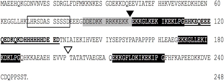
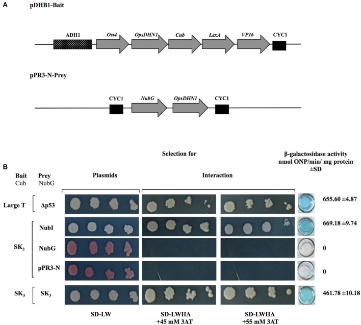
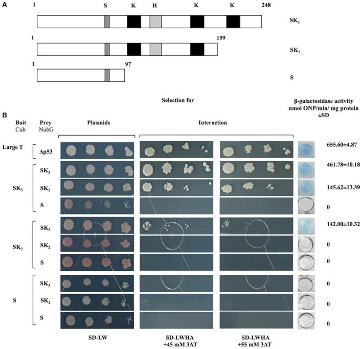
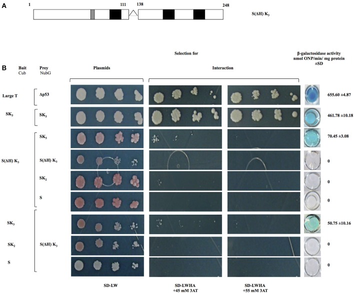
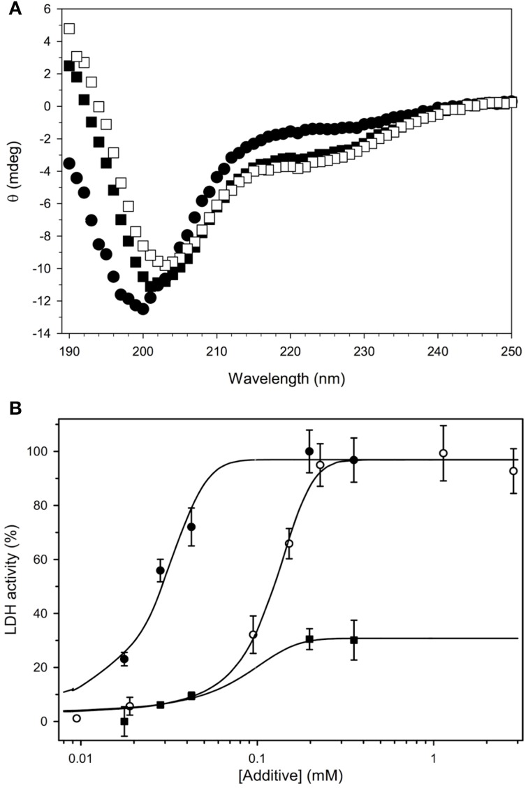
Dehydrins belongs to a large group of highly hydrophilic proteins known as Late Embryogenesis Abundant (LEA) proteins. It is well known that dehydrins are intrinsically disordered plant proteins that accumulate during the late stages of embryogenesis and in response to abiotic stresses; however, the molecular mechanisms by which their functions are carried out are still unclear. We have previously reported that transgenic Arabidopsis plants overexpressing an Opuntia streptacantha SK3 dehydrin (OpsDHN1) show enhanced tolerance to freezing stress. Herein, we show using a split-ubiquitin yeast two-hybrid system that OpsDHN1 dimerizes. We found that the deletion of regions containing K-segments and the histidine-rich region in the OpsDHN1 protein affects dimer formation. Not surprisingly, in silico protein sequence analysis suggests that OpsDHN1 is an intrinsically disordered protein, an observation that was confirmed by circular dichroism and gel filtration of the recombinantly expressed protein. The addition of zinc triggered the association of recombinantly expressed OpsDHN1 protein, likely through its histidine-rich motif. These data brings new insights about the molecular mechanism of the OpsDHN1 SK3-dehydrin.

Keywords: K-segments; SK3-dehydrin; histidine-rich region; homodimer; intrinsically disordered proteins; yeast two-hybrid.


Figures

Figure 1
Figure 1
The Opuntia streptacantha DHN (OpsDHN1) amino acid sequence, and schematic representation of characteristic motifs present in OpsDHN1 protein: S-segment (open box), poly-lysine rich sequence (gray boxes), K-segments (black boxes), and histidine-rich motif (in bold). The end of S version (black triangle), and the end of SK2 version (open triangle); histidine rich region deleted (underlined sequence).
Figure 2
Figure 2
OpsDHN1-OpsDHN1 protein interaction using the split-ubiquitin yeast two-hybrid assay. (A) Schematic representation of OpsDHN1 bait and prey constructs. In the pDHB1 construct, the OpsDHN1 gene was fused at its N-terminal to the Ost4 membrane protein, and its C-terminal to the ubiquitin C-terminal moiety. LexA-VP16, the artificial transcription factor, is fused at the C-terminal end of ubiquitin. This construct is under control of the ADH1 promoter region and CYC1 terminator region. In the pPR3-N-prey construct, the ubiquitin N-terminal was fused to the OpsDHN1 gene. This construct is under control of CYC1 promoter region and CYC1 terminator region. (B) Yeast cells carrying the control system interaction LargeT-Cub/Δp53-NubG, and functional assay of OpsDHN1 SK3 as bait construct using prey control system vectors: NubI, NubG and pPR3-N, and OpsDHN1-Cub/OpsDHN1-NubG interaction. Yeast strains was plated to an OD600 of 0.8, and at serial 10-fold dilutions on semi-selective (SD-LW) and on selective (SD-LWHA) media supplemented with 3-AT (45 and 55 mM). Quantitative β-Galactosidase activity was assayed by hydrolysis of the o-nitrophenyl-b-galactoside (ONPG), as expressed in nmol ONP/min per mg of protein. Data represent the mean ± SD, (n = 3).
Figure 3
Figure 3
Interaction analyses among OpsDHN1 (SK3) and truncated versions (SK2 and S) using the split-ubiquitin yeast two-hybrid assay. (A) Schematic representations of OpsDHN1 derived versions used in the two-hybrid split ubiquitin system. OpsDHN1 SK3 full version; OpsDHN1 SK2 C-terminal truncated version (residues 1–199); OpsDHN1 S short version (residues 1–97). The capital letters indicates characteristic motifs of OpsDHN1 protein: S (S-segment), K (K-segment), and H (histidine-rich motif). (B) Yeast cells carrying the control system interaction LargeT-Cub/Δp53-NubG. Interaction analysis among baits and preys OpsDHN1 derived versions (SK3, SK2, and S). Yeast strains was plated to an OD600 of 0.8, and at serial 10-fold dilutions on semi-selective (SD-LW) and on selective (SD-LWHA) media supplemented with 3-AT (45 and 55 mM). Quantitative β-Galactosidase activity was assayed by hydrolysis of the o-nitrophenyl-b-galactoside (ONPG), as expressed in nmol ONP/min per mg of protein. Data represent the mean ± SD, (n = 3).
Figure 4
Figure 4
Interaction analyses among S(ΔH) K3, OpsDHN1 (SK3), and truncated versions (SK2 and S) using the split-ubiquitin yeast two-hybrid assay. (A) Schematic representations of the OpsDHN1 S(ΔH)SK3 version without histidine-rich motif (residues 1–111:128–248). (B) Yeast cells expressing the control system interaction LargeT-Cub/Δp53-NubG, OpsDHN1 SK3 full-length interaction. Interaction analysis among bait OpsDHN1 S(ΔH) K3-Cub with OpsDHN1-NubG [SK3, S(ΔH) K3, SK2, and S] prey vectors, interaction analysis of the swapped versions in the respective bait and prey vectors. Yeast strains was plated to an OD600 of 0.8, and at serial 10-fold dilutions on semi-selective (SD-LW) and on selective (SD-LWHA) media supplemented with 3-AT (45 and 55 mM). Quantitative β-Galactosidase activity was assayed by hydrolysis of the o-nitrophenyl-b-galactoside (ONPG), as expressed in nmol ONP/min per mg of protein. Data represent the mean ± SD, (n = 3).
Figure 5
Figure 5
Biochemical characterization of recombinant OpsDHN1. (A) The secondary structure content of OpsDHN1 was determined under several conditions. OpsDHN1 alone, filled circles; OpsDHN1 in the presence of 50 mM SDS, filled squares; OpsDHN1 in the presence of 50 mM SDS and 1 mM ZnCl2, open squares. (B) Cryoprotection assay with OpsDHN1. The protection of LDH from freeze-thaw damage was assessed as described in the Materials and Methods. OpsDHN1, filled circles; Vitis riparia K2, open circles; FROST protein, filled squares.

References

    1. Allagulova Ch., R., Gimalov F. R., Shakirova F. M., Vakhitov V. A. (2003). The plant dehydrins: structure and putative functions. Biochemistry Mosc. 68, 945–951 10.1023/A:1026077825584 - DOI - PubMed
    1. Andrews P. (1970). Estimation of molecular size and molecular weights of biological compounds by gel filtration. Methods Biochem. Anal. 18, 1–53 10.1002/9780470110362.ch1 - DOI - PubMed
    1. Battaglia M., Olvera-Carrillo Y., Garciarrubio A., Campos F., Covarrubias A. A. (2008). The enigmatic LEA proteins and other hydrophilins. Plant Physiol. 148, 6–24 10.1104/pp.108.120725 - DOI - PMC - PubMed
    1. Close T. J. (1996). Dehydrins: emergence of a biochemical role of a family of plant dehydration proteins. Physiol. Planta 97, 795–803 10.1111/j.1399-3054.1996.tb00546.x - DOI
    1. Close T. J. (1997). Dehydrins: a commonalty in the response of plants to dehydration and low temperature. Physiol. Planta 100, 291–296 10.1111/j.1399-3054.1997.tb04785.x - DOI

LinkOut - more resources