. 2014 May;5(3):209.
doi: 10.4172/2155-9562.1000209.

T-Lymphocyte Deficiency Exacerbates Behavioral Deficits in the 6-OHDA Unilateral Lesion Rat Model for Parkinson's Disease

Affiliations

T-Lymphocyte Deficiency Exacerbates Behavioral Deficits in the 6-OHDA Unilateral Lesion Rat Model for Parkinson's Disease

Christopher J Wheeler et al. J Neurol Neurophysiol. 2014 May.

Abstract

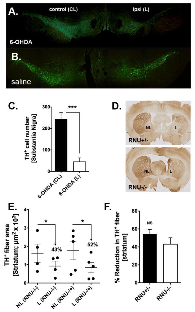
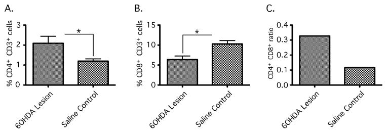
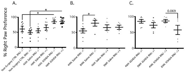
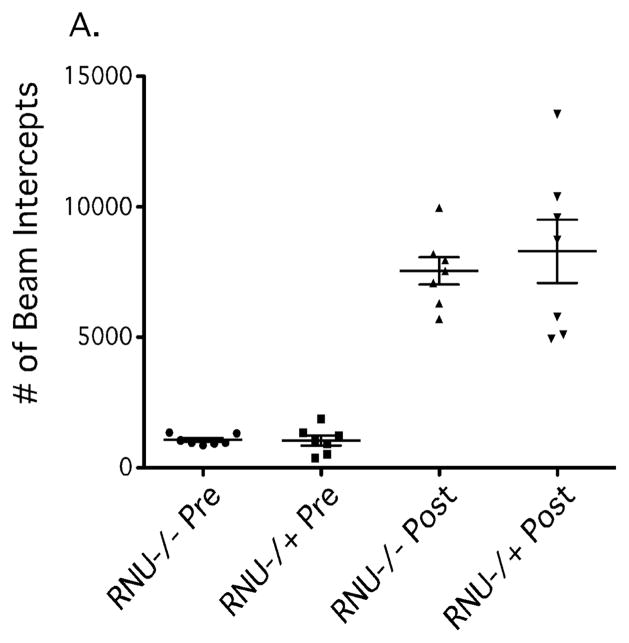
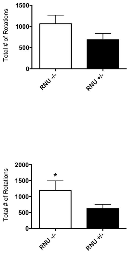
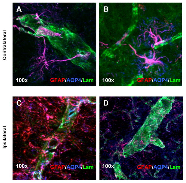
T-lymphocytes have been previously implicated in protecting dopaminergic neurons in the substantianigra from induced cell death. However, the role of T-cells in neurodegenerative models such as Parkinson's disease (PD) has not been fully elucidated. To examine the role of T-lymphocytes on motor behavior in the 6-hydroxydopamine (6-OHDA) unilateral striatal partial lesion PD rat model, we assessed progression of hemi-parkinsonian lesions in the substantia nigra, induced by 6-OHDA striatal injections, in athymic rats (RNU-/-, T-lymphocyte-deficient) as compared to RNU-/+ rats (phenotypically normal). Motor skills were determined by the cylinder and D-amphetamine sulfate-induced rotational behavioral tests. Cylinder behavioral test showed no significant difference between unilaterally lesioned RNU-/- and RNU-/+ rats. However both unilaterally lesioned RNU-/- and RNU-/+ rats favored the use of the limb ipsilateral to lesion. Additionally, amphetamine-induced rotational test revealed greater rotational asymmetry in RNU-/- rats compared to RNU-/+ rats at two- and six-week post-lesion. Quantitative immunohistochemistry confirmed loss of striatal TH-immunopositive fibers in RNU-/- and RNU-/+ rat, as well as blood-brain-barrier changes associated with PD that may influence passage of immune cells into the central nervous system in RNU-/- brains. Specifically, GFAP immunopositive cells were decreased, as were astrocytic end-feet (AQP4) contacting blood vessels (laminin) in the lesioned relative to contralateral striatum. Flow cytometric analysis in 6-OHDA lesioned RNU-/+rats revealed increased CD4+ and decreased CD8+ T cells specifically within lesioned brain. These results suggest that both major T cell subpopulations are significantly and reciprocally altered following 6-OHDA-lesioning, and that global T cell deficiency exacerbates motor behavioral defects in this rat model of PD.


Figures

Figure 1
Figure 1
Tyrosine hydroxylase (TH) immunohistochemistry in the substantia nigra (SN) and striatum. (A) Representative immunofluorescent (IF) microphotographs demonstrating extensive loss of midbrain TH+ dopamine (DA) neuronal cell bodies (green) in the SN of RNU−/+ rats 4 weeks after unilateral 6-hydroxydopamine (6-OHDA) lesion. (B) No significant loss is observed in the SN of RNU−/+ rats following saline injection. (C) Quantitative analysis of TH-immunoreactive DA cells confirms a significant loss in cell number induced by 6-OHDA in the ipsilateral SN (ipsi lesion − L) as compared to the contralateral SN (control − CL) of RNU−/+ rats. (D) Immunohistochemical images indicate a depletion of TH-positivefiber density (brown) in the striatum of both RNU−/+ and RNU−/− rats 8 weeks after 6-OHDA injection (lesion − L,non-lesion − NL). (E) Quantitative analysis of TH+ fiber area confirms a significant reduction of ≈40–50% in striatal fiber density in the injected sites (X + SEM, Y + SEM, respectively). (F) Percent reduction of TH-fiber density after 6-OHDA injections does not differ significantly between RNU−/+ and RNU−/− rats; N = 3–4 rats per group.
Figure 2
Figure 2
Changes in Cerebral T cell populations in the 6-OHDA lesioned RNU−/+ rat brain assessed by Flow cytometry. Striatal and nigral tissue was isolated from RNU−/+ rats 4 weeks after receiving unilateral 6-OHDA toxin or saline as control. Ipsilateral and contralateral to lesion tissue was isolated and dissociated to single cell suspensions for immunocytochemistry to identify CD3+CD4+ and CD3+CD8+ T cells. Cells were than analyzed by flow cytometry and expressed as percent of gated lymphocytes. 6-OHDA tissue showed a significant increase in CD3+CD4+ (%2.091−/+ 0.3538 vs. %1.195 −/+ 0.1160; *, P= 0.0210) immunopositive cells and decrease in CD3+CD8+ (%6.382−/+ 0.8912 vs. %10.27−/+ 0.8661; *, P=0.0034) immunopositive cells as compared to saline injected controls (A+B) and an increase in the ratio of CD4+CD8+ in 6-OHDA RNU−/+ as compared to controls (C). Error bars equal SEM (*, P<0.05).
Figure 3
Figure 3
Athymic rats show attenuated asymmetric forelimb recovery by Cylinder Tests after unilateral 6-OHDA intrastriatal lesion of the nigro-striatal dopaminergic pathway. At 4 and 8 weeks post unilateral 6-OHDA lesion we used the Cylinder test to determine preferential paw. At 4 weeks post lesion RNU−/− and RNU−/+ showed a significant preferential right paw use as compared to non-surgery controls RNU−/−(%60.87 −/+ 9.249 vs. %84.91 −/+ 5.814; *, P = 0.026); RNU−/+ (48.69 −/+ 5.618 vs. 85.94 −/+ 4.685; *, P = 0.0002) (A). At 8 weeks post saline RNU−/− showed significant right paw preference as compared to 4 wks post saline (%54.81 −/+ 6.614 vs. 79.60 −/+ 9.261; *, P = 0.0236) (B). At 8 weeks post 6OHDA lesion RNU −/+ showed a trend towards recovery from right paw preference (%57.94 −/+ 14.49 vs. 85.94 −/+ 4.685; P = 0.069), while RNU−/− shows no significant change in right paw use (%72.54 −/+ 8.286 vs. 84.91 −/+ 5.814; P = 0.2529). Error bars equal SEM (*, P<0.05).
Figure 4
Figure 4
Open Field test of Total Ambulatory and Central locomotor activity in RNU−/− and RNU−/+ after D-amphetamine sulfate administration. In order to assess any potential differences in the metabolism of the drug, D-amphetamine sulfate, and subsequent locomotor activity between RNU−/− and RNU−/+ rats, an Open Field test was conducted prior to surgery (i.e. prior to saline or 6-OHDA injection). We found no significant differences in overall ambulatory (A) during a 120-minute time interval. Error bars equal SEM (*, p=0.05).
Figure 5
Figure 5
Athymic rats exhibit greater D-Amphetamine-induced rotational behavior following unilateral 6-OHDA striatal lesion of the nigro-striatal pathway. Two weeks afterunilateral 6-OHDA lesioned (right) RNU−/− rats show a trend towards increased rotational behavioral ipsilateral to the lesion (right) after d-amphetamine sulphate administration as compared to RNU−/+ rats (1064 −/+ 203.4, N=5 vs. 685.4 −/+ 151.7, N=8; *, P = 0.071) (A). At six weeks post-lesion there was a significant increase in RNU−/− rotational behavior as compared to RNU−/+rats (1188 −/+ 306.5, N=5 vs. 622.8 −/+ 129, N=8; *, P = 0.037) (B). Error bars equal SEM (*, p=0.05).
Figure 6
Figure 6
Immunohistochemistry and blood-brain barrier analysis in 6-OHDA lesioned RNU−/− rats. Laser-scanning confocal microscopy images of triple immunostained tissues using antibodies against GFAP (red), AQP4 (blue) and laminin (green) indicate BBB changes between the contralateral (A and B) and 6-OHDA lesioned brain (C and D) in RNU−/− rats. Localized morphological and protein expression changes suggests differences in the interaction between astrocytes and blood vessels 4 weeks post 6-OHDA lesion between the contralateral and ipsilateral side in RNU−/−. Magnification at 100× indicated direct association of astrocytic end-feet on blood vessels (A and B). However, AQP4 immunoreactivity was diminished and not localized to blood vessels (laminin/green) in the lesioned tissue (C and D).

References

    1. Reynolds AD, Banerjee R, Liu J, Gendelman HE, Mosley RL. Neuroprotective activities of CD4+CD25+ regulatory T cells in an animal model of Parkinson’s disease. J Leukoc Biol. 2007;82:1083–1094. - PubMed
    1. Brochard V, Combadière B, Prigent A, Laouar Y, Perrin A, et al. Infiltration of CD4+ lymphocytes into the brain contributes to neurodegeneration in a mouse model of Parkinson disease. J Clin Invest. 2009;119:182–192. - PMC - PubMed
    1. McGeer PL, Itagaki S, Boyes BE, McGeer EG. Reactive microglia are positive for HLA-DR in the substantia nigra of Parkinson’s and Alzheimer’s disease brains. Neurology. 1988;38:1285–1291. - PubMed
    1. Mogi M, Harada M, Narabayashi H, Inagaki H, Minami M, et al. Interleukin (IL)-1 beta, IL-2, IL-4, IL-6 and transforming growth factor-alpha levels are elevated in ventricular cerebrospinal fluid in juvenile parkinsonism and Parkinson’s disease. Neurosci Lett. 1996;211:13–16. - PubMed
    1. Baba Y, Kuroiwa A, Uitti RJ, Wszolek ZK, Yamada T. Alterations of T-lymphocyte populations in Parkinson disease. Parkinsonism Relat Disord. 2005;11:493–498. - PubMed

LinkOut - more resources