. 2014 Oct 9:2:184.
doi: 10.3389/fpubh.2014.00184. eCollection 2014.

Comparative Study of the Pathological Effects of Western Equine Encephalomyelitis Virus in Four Strains of Culex tarsalis Coquillett (Diptera: Culicidae)

Affiliations

Comparative Study of the Pathological Effects of Western Equine Encephalomyelitis Virus in Four Strains of Culex tarsalis Coquillett (Diptera: Culicidae)

Marco V Neira et al. Front Public Health. .

Abstract

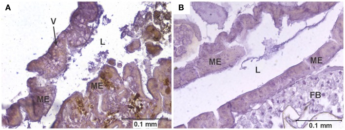
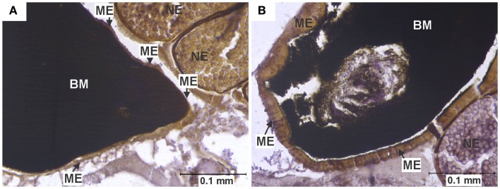
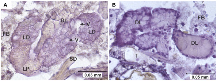
Early reports suggested that mosquito cells infected with arboviruses remain viable and undamaged. However, more recent experimental evidence suggests that arboviral infection of mosquito tissues might indeed result in pathological changes, with potential implications for vector survival and virus transmission. Here, we compare the pathological effects of western equine encephalomyelitis virus (WEEV) infection in four strains of Culex tarsalis previously reported to differ in their competence as WEEV vectors. Pathological effects were observed in cells of the midgut epithelium, salivary glands, and eggs. Cell rounding and sloughing of midgut epithelial cells was associated with those strains reported to be the least susceptible to WEEV infection, whereas midgut necrosis and vacuolation upon infection were associated with strains showing higher susceptibility. Although pathological effects were sporadically observed in infected salivary glands, further studies are required to evaluate their impact on vector competence. Additionally, the potential implications of observed C. tarsalis egg infection with WEEV are discussed.

Keywords: Culex tarsalis; arbovirus; mosquito; pathology; vector competence; western equine encephalomyelitis.


Figures

Figure 1
Figure 1
Vacuolation of midgut epithelium. (A) Section of the posterior midgut of a specimen of the COAV strain, 3 days after ingesting a blood meal containing 5-log PFU of WEEV per 0.1 ml blood. Notice the extensive formation of vacuoles in the cytoplasm. Rusty-brown staining is indicative of a positive immunocytochemical reaction, denoting the presence of WEEV antigen in the tissue. (B) Comparable section of the posterior midgut in an uninfected control. FB, fat body; L, midgut lumen; ME, midgut epithelium; V, vacuole.
Figure 2
Figure 2
Midgut epithelium necrosis. (A) Section of the posterior midgut of a specimen of the HVP strain, 2 days after ingesting a blood meal containing 5-log PFU of WEEV per 0.1 ml blood. Notice how the epithelium has become necrotic, being reduced to a very thin band, with neither discernible cell boundaries nor traces of cytoplasm or organelles. (B) Comparable section of posterior midgut in an uninfected control, where no necrosis is observed. BM, blood meal (in the midgut lumen); ME, midgut epithelium; NE, non-infected egg.
Figure 3
Figure 3
Cell rounding and sloughing (CRS). (A) Anterior midgut section of a specimen of the KNWR strain, 14 days after ingesting a blood meal containing 3-log PFU of WEEV per 0.1 ml blood. Notice how several epithelial cells have sloughed-off into the lumen, and some rounded cells protrude from the tissue. (B) Comparable section of anterior midgut in an uninfected control, where no CRS is observed. FB, Fat body; L, midgut lumen; M, skeletal muscle; ME, midgut epithelium; R, rounded epithelial cell; S, sloughed epithelial cell.
Figure 4
Figure 4
Egg infection and pathology. (A) Longitudinal section of the abdomen of specimen of the COAV strain, 21 days after ingesting a blood meal containing 5-log PFU of WEEV per 0.1 ml blood. Rusty-brown staining is indicative of a positive immunocytochemical reaction, denoting the presence of WEEV antigen in the tissue. Notice the smooth yolk texture observed in infected egg, in contrast with the uniformly granular texture of yolk observed in the neighboring uninfected eggs. (B) Comparable section of the abdomen of a specimen of the COAV strain, showing only normal, uninfected eggs. IE, infected egg; UE, uninfected egg.
Figure 5
Figure 5
Pathology in salivary glands. (A) Salivary gland of a specimen of the HVP strain, 7 days after ingesting a blood meal containing 5-log PFU of WEEV per 0.1 ml blood. Notice the extensive cytoplasmic vacuolation of acinar cells, and the absence of staining indicative of viral antigen. (B) Comparable section of a normal salivary gland from an uninfected control. DL, distal lobe; FB, fat body; LD, lateral-distal lobe; LP, lateral-proximal lobe; SD, salivary duct; T, trachea; V, vacuole.
Figure 6
Figure 6
Frequency of pathological changes in the midgut of C. tarsalis. Cell rounding and sloughing was the only pathology found in uninfected individuals. Signs of necrosis were only found in HVP and COAV strains, consistently at 48 h after infection. COAV, Coachella Valley; CRS, cell rounding and sloughing; HVP, high viremia producer; KNWR, Kern National Wildlife Refuge; WR, WEEV resistant.
Figure 7
Figure 7
Frequency of virus-induced pathology in eggs of C. tarsalis. No significant differences (P > 0.05) in the number of individuals presenting pathological eggs were found either between dose groups or between strains. COAV, Coachella Valley; HVP, high viremia producer; KNWR, Kern National Wildlife Refuge; WR, WEEV resistant.

References

    1. Chamberlain RW, Sudia WD. Mechanism of transmission of viruses by mosquitoes. Annu Rev Entomol (1961) 6:371–90 10.1146/annurev.en.06.010161.002103 - DOI - PubMed
    1. McLintock J. Mosquito-virus relationships of American encephalitides. Annu Rev Entomol (1978) 23:17–37 10.1146/annurev.en.23.010178.000313 - DOI - PubMed
    1. Hardy JL, Houk EJ, Kramer LD, Reeves WC. Intrinsic factors affecting vector competence of mosquitoes for arboviruses. Annu Rev Entomol (1983) 28:229–62 10.1146/annurev.en.28.010183.001305 - DOI - PubMed
    1. Mims CA, Day MF, Marshall ID. Cytopathic effect of Semliki forest virus in the mosquito Aedes aegypti. Am J Trop Med Hyg (1966) 15:775–84 - PubMed
    1. Weaver SC, Scott TW, Lorenz LH, Lerdthusnee K, Romoser WS. Togavirus-associated pathologic changes in the midgut of a natural mosquito vector. J Virol (1988) 62:2083–90 - PMC - PubMed

LinkOut - more resources