Clinical Trial
. 2015 Jul;85(4):616-24.
doi: 10.2319/032414-220.1. Epub 2014 Oct 27.

A pilot clinical study of Class III surgical patients facilitated by improved accelerated osteogenic orthodontic treatments

Affiliations
Clinical Trial

A pilot clinical study of Class III surgical patients facilitated by improved accelerated osteogenic orthodontic treatments

JiaQi Wu et al. Angle Orthod. 2015 Jul.

Abstract

Objective: To evaluate if the improved accelerated osteogenic orthodontics (IAOO) procedure could speed Class III surgical patients' preoperative orthodontic treatment duration and, if yes, to what extent. This study was also designed to determine whether or not an IAOO procedure affects the tooth-moving pattern during extraction space closure.

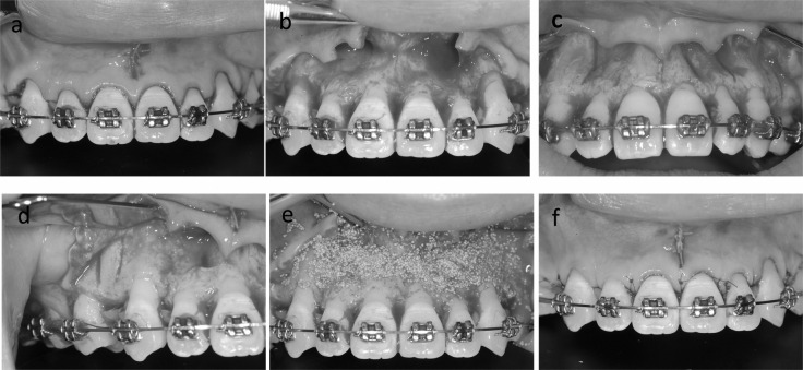
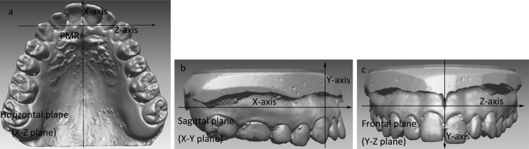
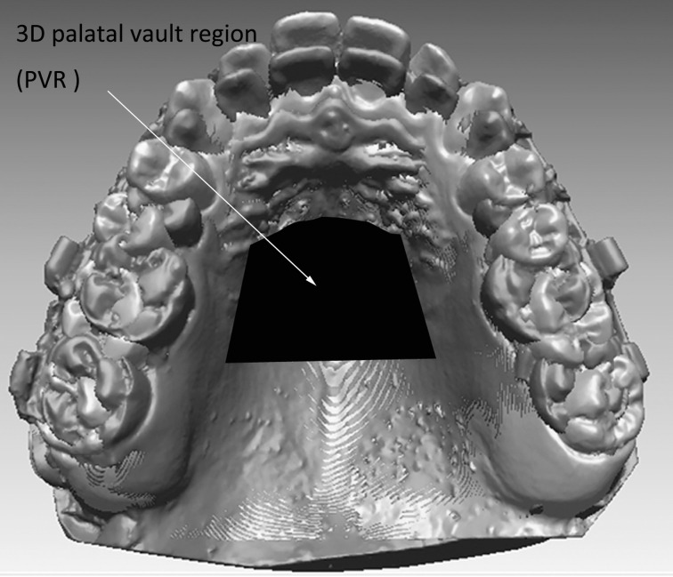
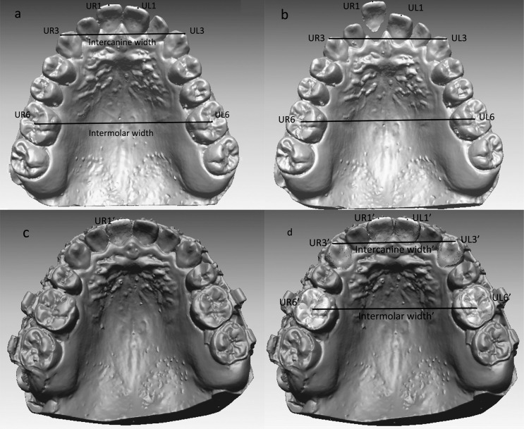
Materials and methods: The samples in this study consisted of 24 Class III surgical patients. Twelve skeletal Class III surgery patients served as an experimental group (group 1) and the others as a control group (group 2). Before treatment, the maxillary first premolars were removed. For group 1, after the maxillary dental arch was aligned and leveled (T2), IAOO procedures were performed in the maxillary alveolar bone. Except for this IAOO procedure in group 1, all 24 patients experienced similar combined orthodontic and orthognathic treatment. Study casts of the maxillary dentitions were made before orthodontic treatment (T1) and after extraction space closure (T3). All of the casts were laser scanned, and the amount of movement of the maxillary central incisor, canine, and first molar, as well as arch widths, were digitally measured and analyzed by using the three-dimensional model superimposition method.

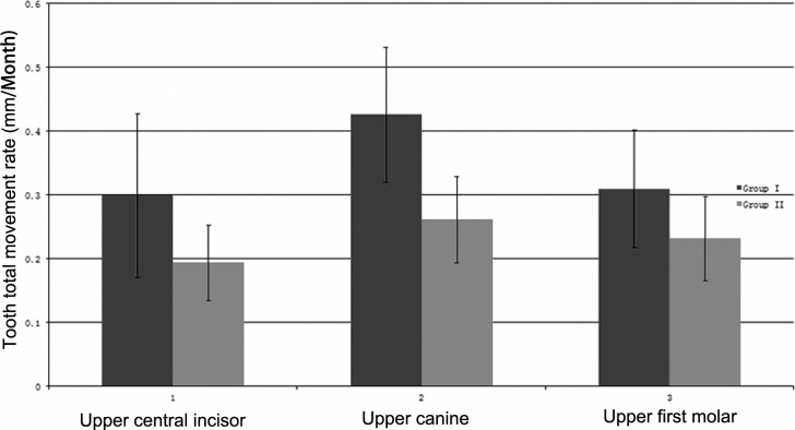
Results: The time durations T3-T2 were significantly reduced in group 1 by 8.65 ± 2.67 months and for T3-T1 were reduced by 6.39 ± 2.00 months (P < .001). Meanwhile, the tooth movement rates were all higher in group 1 (P < .05). There were no significant differences in the amount of teeth movement in the sagittal, vertical, and transverse dimensions between the two groups (P > .05).

Conclusion: The IAOO can reduce the surgical orthodontic treatment time for the skeletal Class III surgical patient by more than half a year on average. The IAOO procedures do not save anchorage.

Keywords: Class III surgical patients; Corticotomy; Improved accelerated osteogenic orthodontics; Preoperative orthodontic treatment duration; Three-dimensional measurement; Tooth movement pattern.


Figures

Figure 1. (a) Sulcular incision performed on the anterior teeth area. (b) Reflection of full-thickness flap. (c, d) Interradicular alveolar corticotomy grooves with piezosurgery. (e) Bone graft material filled the corticotomy area. (f) Flap was sutured.
Figure 1.
(a) Sulcular incision performed on the anterior teeth area. (b) Reflection of full-thickness flap. (c, d) Interradicular alveolar corticotomy grooves with piezosurgery. (e) Bone graft material filled the corticotomy area. (f) Flap was sutured.
Figure 2. The coordinate system. (a) The x-z horizontal plane is the section that includes the junction of inclusive papilla and palatine raphe (PMRJ) and parallel to the occlusal plane constructed by connecting the mesio-buccal cusp tips of the right and left maxillary first molars and the midpoints of the central incisor edges using the best-fitting process. (b) The x-y sagittal plane is the section inclusive of one arbitrary point on the midpalatal suture and the PMRJ and perpendicular to the x-z horizontal plane. (c) The x-z frontal plane is the section inclusive of the PMRJ and perpendicular to both the horizontal and sagittal planes.
Figure 2.
The coordinate system. (a) The x-z horizontal plane is the section that includes the junction of inclusive papilla and palatine raphe (PMRJ) and parallel to the occlusal plane constructed by connecting the mesio-buccal cusp tips of the right and left maxillary first molars and the midpoints of the central incisor edges using the best-fitting process. (b) The x-y sagittal plane is the section inclusive of one arbitrary point on the midpalatal suture and the PMRJ and perpendicular to the x-z horizontal plane. (c) The x-z frontal plane is the section inclusive of the PMRJ and perpendicular to both the horizontal and sagittal planes.
Figure 3. Superimposition of maxillary digital casts of T1 (in blue; please see the online version for color) and T3 (in red) on the three-dimensional palatal vault region.
Figure 3.
Superimposition of maxillary digital casts of T1 (in blue; please see the online version for color) and T3 (in red) on the three-dimensional palatal vault region.
Figure 4. (a) The measuring points on T1 scan (in blue; please see the online version for color). UR1 indicates the midpoint on the edge of maxillary right central incisor; UL1, midpoint on the edge of maxillary left central incisor; UR3, tip of maxillary right canine; UL3, tip of maxillary left canine; UR6, fovea of right maxillary first molar; UL6, fovea of maxillary left first molar; intercanine width, distance between UR3 and UL3; intermolar width, distance between UR6 and UL6. (b) The maxillary right central incisor was detached from the T1 scan. (c) UR1 fixed on the maxillary right central incisor on the T1 scan was transferred into the maxillary right central incisor on the T3 scan (in red) with the three-dimensional image registration technique. (d) The measuring points on the T1 scan were transferred into the corresponding teeth on the T3 scan, respectively.
Figure 4.
(a) The measuring points on T1 scan (in blue; please see the online version for color). UR1 indicates the midpoint on the edge of maxillary right central incisor; UL1, midpoint on the edge of maxillary left central incisor; UR3, tip of maxillary right canine; UL3, tip of maxillary left canine; UR6, fovea of right maxillary first molar; UL6, fovea of maxillary left first molar; intercanine width, distance between UR3 and UL3; intermolar width, distance between UR6 and UL6. (b) The maxillary right central incisor was detached from the T1 scan. (c) UR1 fixed on the maxillary right central incisor on the T1 scan was transferred into the maxillary right central incisor on the T3 scan (in red) with the three-dimensional image registration technique. (d) The measuring points on the T1 scan were transferred into the corresponding teeth on the T3 scan, respectively.
Figure 5. Graphic presentation of total tooth movement rate of group 1 and group 2 from T1 to T3.
Figure 5.
Graphic presentation of total tooth movement rate of group 1 and group 2 from T1 to T3.

References

    1. Phillips C, Proffit WR. Contemporary Treatment of Dentofacial Deformity. St Louis, Mo: Mosby; 2003. Psychosocial aspects of dentofacial deformity and its treatment; p. 69.
    1. Luther F, Morris DO, Hart C. Orthodontic preparation for orthognathic surgery: how long does it take and why? A retrospective study. Br J Oral Maxillofac Surg. 2003;41:401–406. - PubMed
    1. Luther F, Morris DO, Karnezi K. Orthodontic treatment following orthognathic surgery: how long does it take and why? A retrospective study. J Oral Maxillofac Surg. 2007;65:1969–1976. - PubMed
    1. Wilcko TM, William MW, Bissada NF. An evidence-based analysis of periodontally accelerated orthodontic and osteogenic techniques: a synthesis of scientific perspectives. Semin Orthod. 2008;14:305–316.
    1. Hassan AH, Al-Fraidi AA, Al-Saeed SH. Corticotomy-assisted orthodontic treatment: review. Open Dent J. 2010;4:159–164. - PMC - PubMed

Publication types

MeSH terms