. 2015 Jan;95(1):100-12.
doi: 10.1038/labinvest.2014.133. Epub 2014 Oct 27.

Methods for detecting circulating cancer stem cells (CCSCs) as a novel approach for diagnosis of colon cancer relapse/metastasis

Affiliations

Methods for detecting circulating cancer stem cells (CCSCs) as a novel approach for diagnosis of colon cancer relapse/metastasis

Carla Kantara et al. Lab Invest. 2015 Jan.

Abstract

Cancer stem cells (CSCs) are believed to be resistant to currently available therapies and may be responsible for relapse of cancer in patients. Measuring circulating tumor cells (CTCs) in the blood of patients has emerged as a non-invasive diagnostic procedure for screening patients who may be at high risk for developing metastatic cancers or relapse of the cancer disease. However, accurate detection of CTCs has remained a problem, as epithelial-cell markers used to date are not always reliable for detecting CTCs, especially during epithelial-mesenchymal transition. As CSCs are required to initiate metastatic tumors, our goal was to optimize and standardize a method for identifying circulating CSCs (CCSCs) in patients, using established CSC markers. Here, we report for the first time the detection of CCSCs in the blood of athymic nude mice, bearing metastatic tumors, and in the blood of patients positive for colonic adenocarcinomas. Using a simple and non-expensive method, we isolated a relatively pure population of CSCs (CD45-/CK19+), free of red blood cells and largely free of contaminating CD45+ white blood cells. Enriched CCSCs from patients with colon adenocarcinomas had a malignant phenotype and co-expressed CSC markers (DCLK1/LGR5) with CD44/Annexin A2. CSCs were not found in the blood of non-cancer patients, free of colonic growths. Enriched CCSCs from colon cancer patients grew primary spheroids, suggesting the presence of tumor-initiating cells in the blood of these patients. In conclusion, we have developed a novel diagnostic assay for detecting CSCs in circulation, which may more accurately predict the risk of relapse or metastatic disease in patients. As CSCs can potentially initiate metastatic growths, patients positive for CCSCs can be treated with inhibitory agents that selectively target CSCs, besides conventional treatments, to reduce the risk of relapse/metastatic disease for improving clinical outcomes.


Conflict of interest statement

Disclosures: No conflict of interest to report.

Figures

Figure 1
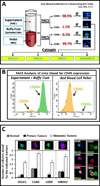
Figure 1. Detection of metastatic circulating tumor cells in blood of athymic nude mice
A) Diagnostic scheme for isolating nucleated epithelial cells from the blood of mice. Three samples of blood/mouse group were collected and analyzed by FACSorting for percent cells positive or negative for CD45 in the supernatant/buffy coat and red blood cell layers. CD45− cells isolated from the supernatant/buffy coat layer are stained by IF staining for the indicated markers. B) Representative data from FACSorting of the cells into CD45+/− populations, showing forward scatter plots for the two fractions of mouse plasma from GroupIII mice. C) Bar graphs depicting percent cells positive for the specific markers/mL blood, collected from all 3 groups. Images of CD45− cells stained by IF for DCLK1/ANXA2/CD44/LGR5 markers are shown above the bar graphs and co-expression is depicted as merged images on the right. Mean±SEM of data obtained from ~10 slides/3 samples/group. *=P< 0.05 versus normal values.
Figure 2
Figure 2. Presence of CD44+/CD45− cells in the blood of patients with colon-adenocarcinomas
Bar graphs illustrating: A) Number of recovered CD45+ cells/mL in the blood of patients, who were either free of colonic growths (normal), or positive for Adenocarcinomas (AdCA); IF images of remaining CD45+ cells collected in the final elution are shown above the bar graphs. B) Number of CD44+ cells/mL in blood of normal vs AdCA patients. C) Number of cells co-localizing for CD45/CD44 per mL of blood. Representative IF staining of circulating cells stained with indicated markers are shown above bar graphs, after negative selection of the samples. D) Number of CD44+ cells/mL, which do not co-localize with CD45 in blood of normal vs AdCA patients; representative IF images are shown above bar graphs. Each bar graph=mean ±SEM of data from 5–7 patients analyzed in triplicate as described in Methods. *=P< 0.05 versus normal values.
Figure 3
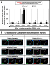
Figure 3. A sub-population of Leukocytes is also positive for DCLK1, ANXA2 and CD44
A) Blood from normal and AdCA patients were processed with the EasySep™ kit, catalog #18289 Cells collected after final elution, were cytospun onto glass slides, followed by IF staining for indicated markers. Based on the staining results, number of cells positive for the indicated markers was determined/mL blood, as shown in the bar graphs. B) Representative IF images showing co-expression of DCLK1/CD45, CD44/CD45 and ANXA2/CD45. Images were taken at 10x magnification and a single cell image was magnified and is presented in an inset. (n=7 normal samples and n=7 AdCA samples).
Figure 4
Figure 4. Circulating cancer stem cells (CCSCs) in the blood of patients
A) Representative IF images of circulating cancer stem cells, positive for indicated markers. Images were digitally enhanced, as shown by dotted arrows. B) Bar graphs illustrating number of cells/mL, positive for indicated markers, excluding CD45+ cells. (n=7 normal samples and n=7 AdCA samples).
Figure 5
Figure 5. Co-expression of CSC markers (DCLK1/LGR5) with AnxA2/PG/CD44/EpCAM/CK19 by CCSCs in the blood of colon cancer patients
A) Co-expression (IF staining) of DCLK1+/LGR5+ cells with either EpCAM, CD44 or ANAX2 in AdCA samples; white arrows depict stained cells. B) Co-expression of DCLK1/PG or ANXA2/PG in CCSCs. C) CCSCs co-expressing CK19 with either ANXA2, DCLK1 or LGR5. Merged images are enhanced in the inset. D) Bar graphs illustrating number of CCSC cells co-expressing indicated markers/mL blood. (n=7 normal samples and n=7 AdCA samples).
Figure 6
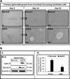
Figure 6. Formation of spheroids, in vitro, from circulating tumor cells, isolated from AdCA patients
A) Representative Images of spheroids formed from enriched circulating epithelial cells, isolated from blood of normal and AdCA patients. Images were taken at 4x and 40x magnification at days 3, 14 and 25 after seeding the cells. Bi) Western blot analysis demonstrating increased expression of LGR5 and DCLK1 in spheroids from CTCs isolated from a representative AdCA patient, compared to that obtained from a representative normal patient. Bii) Bar graphs demonstrating percent change in ratio of target proteins to β-actin. (n=3 normal patient samples; n=3 AdCA patient samples).

References

    1. Siegel R, Desantis C, Jemal A. Colorectal cancer statistics, 2014. CA Cancer J Clin. 2014;64:104–117. - PubMed
    1. Ning X, Shu J, Du Y, et al. Therapeutic strategies targeting cancer stem cells. Cancer Biol Ther. 2013;14:295–303. - PMC - PubMed
    1. Riethdorf S, Wikman H, Pantel K. Review: Biological relevance of disseminated tumor cells in cancer patients. Int J Cancer. 2008;123:1991–2006. - PubMed
    1. Maheswaran S, Haber DA. Circulating tumor cells: a window into cancer biology and metastasis. Curr Opin Genet Dev. 2010;20:96–99. - PMC - PubMed
    1. King MR. Rolling in the deep: therapeutic targeting of circulating tumor cells. Front Oncol. 2012;2:184. - PMC - PubMed

Publication types

Substances