. 2014 Nov 15;5(21):10596-606.
doi: 10.18632/oncotarget.2517.

Angiomatous meningiomas have a distinct genetic profile with multiple chromosomal polysomies including polysomy of chromosome 5

Affiliations

Angiomatous meningiomas have a distinct genetic profile with multiple chromosomal polysomies including polysomy of chromosome 5

Malak S Abedalthagafi et al. Oncotarget. .

Abstract

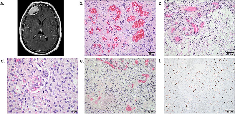
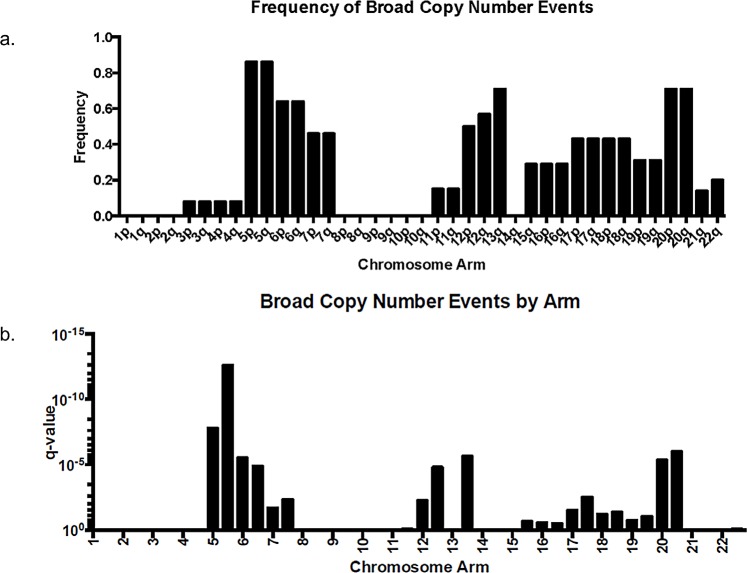
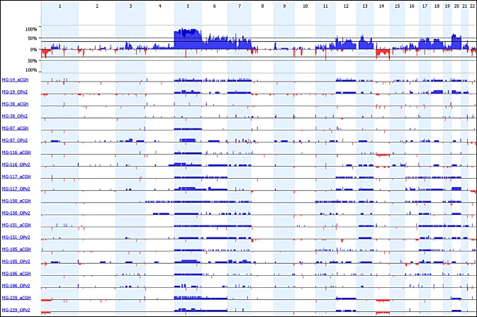
Meningiomas are a diverse group of tumors with a broad spectrum of histologic features. There are over 12 variants of meningioma, whose genetic features are just beginning to be described. Angiomatous meningioma is a World Health Organization (WHO) meningioma variant with a predominance of blood vessels. They are uncommon and confirming the histopathologic classification can be challenging. Given a lack of biomarkers that define the angiomatous subtype and limited understanding of the genetic changes underlying its tumorigenesis, we compared the genomic characteristics of angiomatous meningioma to more common meningioma subtypes. While typical grade I meningiomas demonstrate monosomy of chromosome 22 or lack copy number aberrations, 13 of 14 cases of angiomatous meningioma demonstrated a distinct copy number profile--polysomies of at least one chromosome, but often of many, especially in chromosomes 5, 13, and 20. WHO grade II atypical meningiomas with angiomatous features have both polysomies and genetic aberrations characteristic of other atypical meningiomas. Sequencing of over 560 cancer-relevant genes in 16 cases of angiomatous meningioma showed that these tumors lack common mutations found in other variants of meningioma. Our study demonstrates that angiomatous meningiomas have distinct genomic features that may be clinically useful for their diagnosis.


Conflict of interest statement

Conflict of interest

S. Santagata is a consultant for Bayesian Diagnostics. There are no potential conflicts of interest disclosed by the other authors.

Figures

Figure 1
Figure 1. aCGH of WHO grade I meningioma and angiomatous meningioma (WHO grade I and II)
Plot of copy number gains (blue) and losses (red) for 14 angiomatous meningioma samples (top) and 12 non-angiomatous WHO grade I meningiomas (bottom) that were analyzed using 1×1M Agilent SurePrint G3 Human CGH Microarray chips containing 963,029 probes with 2.1 kb overall median probe spacing and a 1.8 kb probe spacing across the human genome (Oncocopy). Cases MG-19 through MG-229 were analyzed as clinical cases in a CLIA environment by the Center for Advanced Molecular Diagnostics (CAMD) at Brigham and Women's Hospital, and MG-150 through MG-186 were analyzed as research cases, also by the CAMD. All non-angiomatous cases were analyzed for clinical diagnosis. Chromosome numbers are displayed horizontally (even numbers are listed at the top and odd numbers are listed at the bottom). Sex chromosomes were not analyzed or displayed. The segmented aCGH data was plotted using the aberration-plot function of the copy number and Bioconductor packages in R (limits −2,2).
Figure 2
Figure 2. Diversity of morphologies of WHO grade I angiomatous meningioma with polysomies
H&E stained sections demonstrate that while some angiomatous meningiomas have thick hyalinized blood vessels (a-c), others have a mixed profile of capillary-sized blood vessels admixed with hyalinized blood vessels (d), or only predominantly capillary-sized vessels (e, f).
Figure 3
Figure 3. Features of a WHO grade II atypical meningioma with angiomatous features
(a) Axial T1-weighted gadolinium-enhanced MRI of patient MG-229 shows a 4.6 cm right frontal convexity meningioma that extends to the orbital roof, with moderate adjacent vasogenic edema and mild effacement of the anterior horn of the right lateral ventricle. H&E stained sections reveal (b) prominent thick- and thin-walled blood vessels with (c) microcystic change, (d) numerous mitoses, (e) and foci of necrosis, and (f) immunohistochemistry shows an elevated MIB-1 proliferative index. Scale bars and measurements are shown for each image.
Figure 4
Figure 4. Frequency and significance of broad (arm-level) copy number aberrations in 14 angiomatous meningiomas by aCGH
(a.) Frequency (% of the 14 cases having the event) and (b.) q-value data as calculated by GISTIC 2.0 [38] from the 14 angiomatous meningioma cases presented in Figure 1.
Figure 5
Figure 5. Copy number analysis from exon sequencing data of 560 cancer genes
The frequency of gains and losses is shown in the upper panel. The copy number profile from each individual tumor sample is shown in the lower panel (blue – gain; red – loss).
Figure 6
Figure 6. Comparison of copy number analysis from exon sequencing data and high-resolution aCGH data
Ten angiomatous meningiomas were characterized by both exon sequencing (OPv2) and aCGH. The plot displays the copy number changes from the orthogonal analytical approaches (blue - gain; red - loss).

References

    1. Louis DN, Ohgaki H, Wiestler OD, Cavenee WK, Burger PC, Jouvet A, Scheithauer BW, Kleihues P. The 2007 WHO classification of tumours of the central nervous system. Acta Neuropathol. 2007;114(2):97–109. - PMC - PubMed
    1. Wellenreuther R, Kraus JA, Lenartz D, Menon AG, Schramm J, Louis DN, Ramesh V, Gusella JF, Wiestler OD, von Deimling A. Analysis of the neurofibromatosis 2 gene reveals molecular variants of meningioma. Am J Pathol. 1995;146(4):827–832. - PMC - PubMed
    1. Trofatter JA, MacCollin MM, Rutter JL, Murrell JR, Duyao MP, Parry DM, Eldridge R, Kley N, Menon AG, Pulaski K, et al. A novel moesin-, ezrin-, radixin-like gene is a candidate for the neurofibromatosis 2 tumor suppressor. Cell. 1993;72(5):791–800. - PubMed
    1. Ruttledge MH, Sarrazin J, Rangaratnam S, Phelan CM, Twist E, Merel P, Delattre O, Thomas G, Nordenskjold M, Collins VP, et al. Evidence for the complete inactivation of the NF2 gene in the majority of sporadic meningiomas. Nat Genet. 1994;6(2):180–184. - PubMed
    1. Choy W, Kim W, Nagasawa D, Stramotas S, Yew A, Gopen Q, Parsa AT, Yang I. The molecular genetics and tumor pathogenesis of meningiomas and the future directions of meningioma treatments. Neurosurg Focus. 2011;30(5):E6. - PubMed

Publication types

Substances

LinkOut - more resources